Review
. 2016 Mar 3;10(1-2):208-25.
doi: 10.1080/19336918.2016.1148866. Epub 2016 Mar 2.

Neutrophil migration into the placenta: Good, bad or deadly?

Affiliations
Review

Neutrophil migration into the placenta: Good, bad or deadly?

Stavros Giaglis et al. Cell Adh Migr. .

Abstract

Almost 2 decades have passed since the discovery that pregnancy is associated with a basal inflammatory state involving neutrophil activation, and that this is more overt in cases with preeclampsia, than in instances with sepsis. This pivotal observation paved the way for our report, made almost a decade ago, describing the first involvement of neutrophil extracellular traps (NETs) in a non-infectious human pathology, namely preeclampsia, where an abundance of these structures were detected directly in the placental intervillous space. Despite these remarkable findings, there remains a paucity of interest among reproductive biologists in further exploring the role or involvement of neutrophils in pregnancy and related pathologies. In this review we attempt to redress this deficit by highlighting novel recent findings including the discovery of a novel neutrophil subset in the decidua, the interaction of placental protein 13 (PP13) and neutrophils in modulating spiral artery modification, as well as the use of animal model systems to elucidate neutrophil function in implantation, gestation and parturition. These model systems have been particularly useful in identifying key components implicated in recurrent fetal loss, preeclampsia or new signaling molecules such as sphingolipids. Finally, the recent discovery that anti-phospolipid antibodies can trigger NETosis, supports our hypothesis that these structures may contribute to placental dysfunction in pertinent cases with recurrent fetal loss.

Keywords: animal model; neutrophil extracellular traps (NETs); parturition; preeclampsia; pregnancy; recurrent fetal loss.


Figures

Figure 1.
Figure 1.
Sequential steps of neutrophil recruitment from the vasculature to the tissue. Two possible mechanisms of transmigration are described: (a) paracellular - between endothelial cells; and (b) transcellular - through endothelial cells. Major groups of adhesion molecules are marked. Rolling depends mostly on selectins, whereas adhesion, crawling and transmigration depend on integrin interactions. Chemokines lining the lumen of the vascular endothelium activate rolling neutrophils, thus inducing conformational changes of the integrins on the surface of the neutrophils and facilitating the subsequent events. Crawling neutrophils follow the chemokine gradient along the endothelium, which leads them to the preferential sites of transmigration. Figure adapted from ref.
Figure 2.
Figure 2.
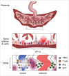
Immune diversion model, spiral artery modification and PP13 Upper panel: The hemochorial human placenta is nourished by maternal blood that is injected into the intervillus space via the uterine spiral arterioles (red decidual vessels). Products of syncytiotrophoblast secretion are released into the intervillus space and, along with blood, are returned to the maternal circulation through the decidual basalis veins (blue decidual vessels). Middle panel: Decidual veins are filled with placental protein 13 (PP13) while PP13 and associated neutrophils transudate to the region. Lower panel: PP13 shows intense deposition consistent with early and active ZONE formation, and other areas of end-stage ZONEs. Neutrophils follow an inverse pattern with the least intense staining in the early ZONEs and the most intense in the endstage ZONEs. Combining this data suggests that syncytiotrophoblast-secreted PP13 exits the intervillus space via the decidual basalis veins (blue) where it binds to the endothelial cells, traverses the veins to be deposited into the surrounding decidua, precipitates, and induces a ZONE consisting of activated T cells, macrophages, and neutrophils. At the same time, invasive trophoblasts migrate to and invade the maternal spiral arterioles (red) without interference from potentially cytotoxic elements of maternal immune surveillance. Figure adapted from ref.
Figure 3.
Figure 3.
Mechanism of antiphospholipid (aPL) antibody–induced fetal damage. aPL antibodies are directed to the placenta where they activate the classical pathway of the complement cascade which leads to the expression of potent anaphylatoxins, C5a in particular. C5a is a neutrophil, monocyte and platelet activator, which furthermore stimulates the release of inflammatory mediators, including reactive oxygen species (ROS), proteolytic enzymes, histones, cytokines and chemokines, as well as additional complement and coagulation factors. Tissue factor (TF) expression on monocytes enhances the release of antiangiogenic molecule sFlt-1. sFlt-1 impairs trophoblast proliferation, reduces placental blood flow, induces oxidative stress, and increases TF expression on trophoblasts. This creates a proinflammatory amplification loop at sites of leukocyte infiltration that generates additional C5a. This results in enforced neutrophil influx, inflammation within the placenta, and ultimately, fetal injury. Either fetal growth restriction or even death in utero ensues depending on the extent of the damage. PMN: neutrophil, Mϕ: monocyte/macrophage. Figure adapted from ref. and ref.
Figure 4.
Figure 4.
Tumor associated granulocytes. Chemokines expressed by tumor cells and tumor-associated macrophages (TAM) promote the recruitment of circulating neutrophils into the tumors. Neutrophils promote genetic instability, possibly through generation of ROS and stimulate angiogenesis through the production of matrix metalloproteinase 9 (MMP9) and vascular endothelial growth factor (VEGF). Transforming growth factorβ (TGFβ) forces neutrophils to obtain a polarized, pro-tumoral N2 phenotype, which is characterized by high levels of arginase production. On the other hand, inhibition of TGFβ promotes neutrophil maturation toward an N1 phenotype. This is associated with higher cytotoxic activity, higher capacity to generate H2O2, higher expression of tumor necrosis factor a (TNFα) and lower expression of arginase and intercellular adhesion molecule 1 (ICAM1), CD8+ T cell activation increases in the presence of N1 neutrophils, which leads to an effective antitumor effect. Figure adapted from ref.
Figure 5.
Figure 5.
Contribution of PMNs to parturition. Neutrophils have multifaceted roles during parturition, either at term or preterm, and are attracted from the systemic circulation to the uterus by a process which is driven by IL-8 (top right) where they release cytokines and MMPs to contribute to labor and post-partum wound sealing and healing. PMNs infiltrate the human cervix only postpartum, again by chemotaxis to IL-8 and CXCL1 and play rather an important role in post-partum tissue repair (bottom right). PMNs infiltrate the human myometrium during term labor by similar conditions, i.e., cyto- and chemokine gradient-driven (bottom left). PMN numbers rise in the human chorioamniotic membranes modestly during term labor, which is consistent with the relatively low increase in the expression of IL-8 in the chorioamniotic membranes and the choriodecidua following term labor. PMNs are recruited in large numbers into the chorioamniotic membranes upon infection and inflammation (i.e., histological chorioamnionitis (top left). Figure adapted from ref.

References

    1. Borregaard N. Neutrophils, from marrow to microbes. Immunity 2010; 33:657-70; PMID:21094463; http://dx.doi.org/10.1016/j.immuni.2010.11.011 - DOI - PubMed
    1. Mocsai A. Diverse novel functions of neutrophils in immunity, inflammation, and beyond. J Exp Med 2013; 210:1283-99; PMID:23825232; http://dx.doi.org/10.1084/jem.20122220 - DOI - PMC - PubMed
    1. Hahn S, Giaglis S, Chowdhury CS, Hosli I, Hasler P. Modulation of neutrophil NETosis: interplay between infectious agents and underlying host physiology. Semin Immunopathol 2013; 35:439-53; PMID:23649713; http://dx.doi.org/10.1007/s00281-013-0380-x - DOI - PMC - PubMed
    1. Lande R, Gregorio J, Facchinetti V, Chatterjee B, Wang YH, Homey B, Cao W, Wang YH, Su B, Nestle FO, et al.. Plasmacytoid dendritic cells sense self-DNA coupled with antimicrobial peptide. Nature 2007; 449:564-9; PMID:17873860; http://dx.doi.org/10.1038/nature06116 - DOI - PubMed
    1. Lande R, Ganguly D, Facchinetti V, Frasca L, Conrad C, Gregorio J, Meller S, Chamilos G, Sebasigari R, Riccieri V, et al.. Neutrophils activate plasmacytoid dendritic cells by releasing self-DNA-peptide complexes in systemic lupus erythematosus. Sci Transl Med 2011; 3:73ra19; PMID:21389263; http://dx.doi.org/10.1126/scitranslmed.3001180 - DOI - PMC - PubMed

LinkOut - more resources