. 2016 Apr 25;7(2):203-15.
doi: 10.1080/19491034.2016.1157674. Epub 2016 Mar 2.

Reproduction of the FC/DFC units in nucleoli

Affiliations

Reproduction of the FC/DFC units in nucleoli

Evgeny Smirnov et al. Nucleus. .

Abstract

The essential structural components of the nucleoli, Fibrillar Centers (FC) and Dense Fibrillar Components (DFC), together compose FC/DFC units, loci of rDNA transcription and early RNA processing. In the present study we followed cell cycle related changes of these units in 2 human sarcoma derived cell lines with stable expression of RFP-PCNA (the sliding clamp protein) and GFP-RPA43 (a subunit of RNA polymerase I, pol I) or GFP-fibrillarin. Correlative light and electron microscopy analysis showed that the pol I and fibrillarin positive nucleolar beads correspond to individual FC/DFC units. In vivo observations showed that at early S phase, when transcriptionally active ribosomal genes were replicated, the number of the units in each cell increased by 60-80%. During that period the units transiently lost pol I, but not fibrillarin. Then, until the end of interphase, number of the units did not change, and their duplication was completed only after the cell division, by mid G1 phase. This peculiar mode of reproduction suggests that a considerable subset of ribosomal genes remain transcriptionally silent from mid S phase to mitosis, but become again active in the postmitotic daughter cells.

Keywords: FC/DFC units; cell cycle; nucleolus; rDNA; replication.


Figures

Figure 1.
Figure 1.
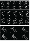
Cell lines with stable expression of fusion proteins. (A) GFP-RPA43 and RFP-PCNA. (B) GFP-fibrillarin and RFP-PCNA. (A) and (B) show one cell passing through G1 and several stages of S phase. Both RPA43 and fibrillarin signals appear as nucleolar beads. Scale bars: (A,B): 5 um
Figure 2.
Figure 2.
FC/DFC units (arrows) in the nucleolus of a cell stably expressing GFP-RPA43. The units are surrounded by the nucleolar granular component (stars). NM – nuclear membrane. Scale bar: 1 μm.
Figure 3.
Figure 3.
CLEM analysis of FC/DFC units. (A, B) Cells on glass-bottom dish supplied with grid; region of interest is framed. (C) The cell selected in (B); a confocal image showing expression of GFP-RPA43 (green) and RFP-PCNA (red). (D) An optical section of the same cell in green channel. (E, F) Ultrathin sections corresponding to the optical section in (D). The marked nucleolar beads in (D) correspond to FC/DFC units in (E) and (F) (framed numbers 1 through 7). Scale bars: 2 μm
Figure 4.
Figure 4.
Short term observations of FC/DFC units during early S phase. Projections of confocal z-stacks representing fragments of the cell nuclei. (A) Disappearing signal (arrows) in the cell expressing GFP-RPA43. Arrowheads show putative re-appearance of a vanished unit (arrowheads). (B,C) Proliferation of the units in the cells expressing GFP-RPA43 (B) or GFP-fibrillarin. (C) The new units appear after splitting of previously existing foci (arrows), or emerge de novo (arrowheads). Scale bars: (A) 500 nm; (B) 500 nm; (C) 3 μm
Figure 5.
Figure 5.
NORs and nucleolar necklaces in the cells expressing GFP-RPA43. Projections of confocal z-stacks. (A) After telophase, each mitotic NOR (arrow) gradually unfolds into a necklace (arrowheads). Nucleoli are formed from one or more necklaces. Emerging nuclei of the daughter cells are outlined in the first frame representing telophase. (B) Interphase cell treated with calyculin A for 1 h. A part of cell nucleus is shown. Nucleolar necklaces (arrowheads) gradually shrink into NORs (arrow). Scale bars: (A) 2 µm, (B) 3 μm
Figure 6.
Figure 6.
Dynamics of the number of FC/DFC units in the course of cell cycle. (A) Counts of the GFP-RPA43 positive units in individual cells. (B) Averaged data for both GFP-RPA43 and GFP-fibrillarin positive units. The values are presented as proportions to the numbers at G1 phase. In (A) and (B) the number of units increases twice: between G1 and mid S phase by cca 70%; in the daughter postmitotic cells at G1 by cca 15%. (C,D,E) Nucleolar beads stained with anti pol I antibody in synchronized HeLa and LEP cells. (C) Counts of the beads show incomplete duplication (increase by 62% in HeLa and by 75% in LEP cells) between G1 and late S (stages S3 and S4). (D,E): pol I (green) and incorporated EdU (red) in a HeLa (D) and LEP (E) cell. The replication signals correspond to S1 stage. Scale bar: 2 μm
Figure 7.
Figure 7.
A hypothesis of reproduction of the transcriptionally active ribosomal genes in the cell cycle. The genes are shown as small circles assembled in groups (NORs). The top row represents progress of a cell from G1 to metaphase. After S phase the genes (small circles assembled in groups) are duplicated, but not all of them are active. During mitosis, when transcription is suspended, the former active genes remain competent. In the daughter postmitotic cells (bottom row), during early G1 phase, the former active and the former silent genes become active. Thus the stable state of activity is maintained therough the consecutive cell cycles. The diagram does not show the permanently inactive genes and NORs which are not associated with pol I.

References

    1. Henderson AS, Warburton D, Atwood KC. Location of ribosomal DNA in the human chromosome complement. Proc Natl Acad Sci USA 1972; 69:3394-8; PMID:4508329; http://dx.doi.org/10.1073/pnas.69.11.3394 - DOI - PMC - PubMed
    1. Long EO, Dawid IB. Repeated genes in eukaryotes. Annu Rev Biochem 1980; 49:727-64; PMID:6996571; http://dx.doi.org/10.1146/annurev.bi.49.070180.003455 - DOI - PubMed
    1. Puvion-Dutilleul F, Bachellerie JP, Puvion E. Nucleolar organization of HeLa cells as studied by in situ hybridization. Chromosoma 1991; 100:395-409; PMID:1893795; http://dx.doi.org/10.1007/BF00337518 - DOI - PubMed
    1. Raska I. Oldies but goldies: searching for Christmas trees within the nucleolar architecture. Trends Cell Biol 2003; 13:517-25; PMID:14507479; http://dx.doi.org/10.1016/j.tcb.2003.08.003 - DOI - PubMed
    1. Raska I, Shaw PJ, Cmarko D. New insights into nucleolar architecture and activity. Int Rev Cytol 2006a; 255:177-235; http://dx.doi.org/10.1016/S0074-7696(06)55004-1 - DOI - PubMed

Publication types