. 2016 May;34(5):1239-50.
doi: 10.1002/stem.2351. Epub 2016 Mar 28.

Genetic Tagging During Human Mesoderm Differentiation Reveals Tripotent Lateral Plate Mesodermal Progenitors

Affiliations

Genetic Tagging During Human Mesoderm Differentiation Reveals Tripotent Lateral Plate Mesodermal Progenitors

Chee Jia Chin et al. Stem Cells. 2016 May.

Abstract

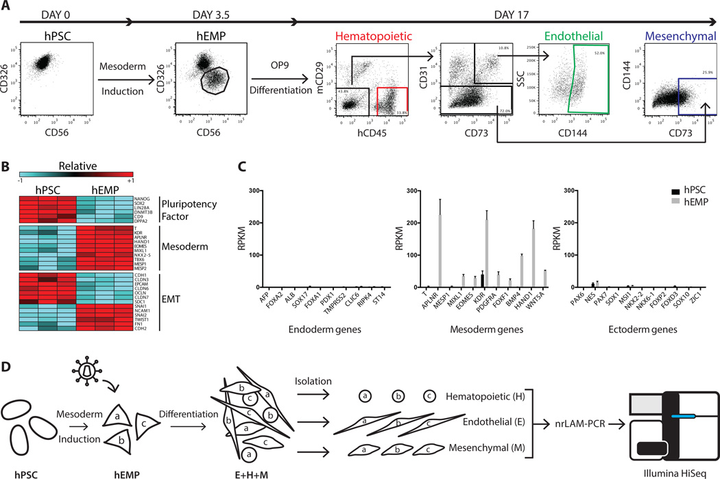
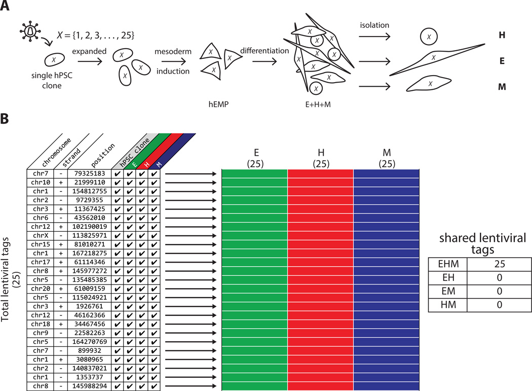
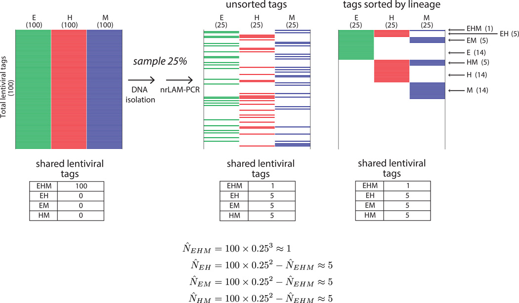
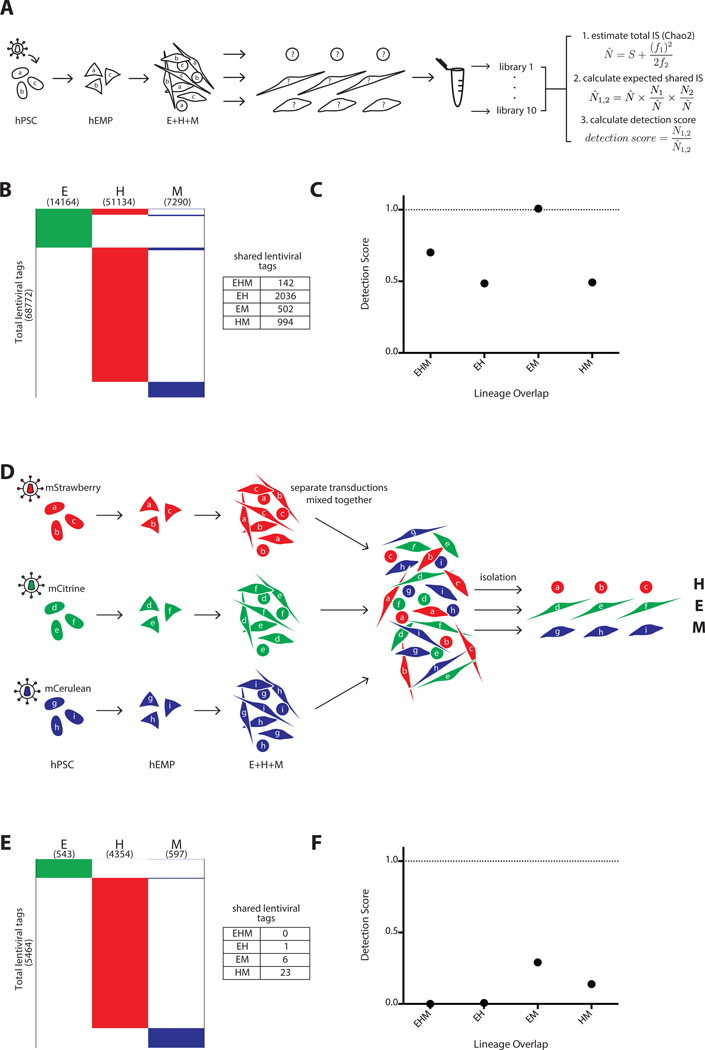
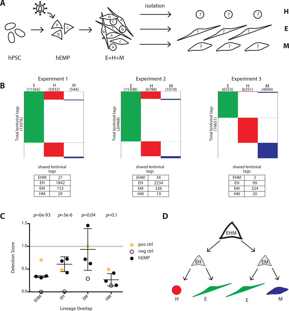
Although clonal studies of lineage potential have been extensively applied to organ specific stem and progenitor cells, much less is known about the clonal origins of lineages formed from the germ layers in early embryogenesis. We applied lentiviral tagging followed by vector integration site analysis (VISA) with high-throughput sequencing to investigate the ontogeny of the hematopoietic, endothelial and mesenchymal lineages as they emerge from human embryonic mesoderm. In contrast to studies that have used VISA to track differentiation of self-renewing stem cell clones that amplify significantly over time, we focused on a population of progenitor clones with limited self-renewal capability. Our analyses uncovered the critical influence of sampling on the interpretation of lentiviral tag sharing, particularly among complex populations with minimal clonal duplication. By applying a quantitative framework to estimate the degree of undersampling we revealed the existence of tripotent mesodermal progenitors derived from pluripotent stem cells, and the subsequent bifurcation of their differentiation into bipotent endothelial/hematopoietic or endothelial/mesenchymal progenitors. Stem Cells 2016;34:1239-1250.

Keywords: Clonal tracking; Hematopoiesis; Lentiviral vectors; Lineage tracing; Mesoderm; Pluripotent stem cells; Vector integration site analysis.


Conflict of interest statement

of Potential Conflicts of Interest The authors indicate no potential conflicts of interest.

Figures

Figure 1
Figure 1
Generation of a human embryonic mesodermal progenitor population from hPSC with hematopoietic, endothelial, and mesenchymal potential. (A): Flow cytometry analysis of EPCAM/CD326 and NCAM/CD56 expression in undifferentiated hPSC and hEMP generated after 3.5 days of sequential morphogen induction with Activin A, BMP4, VEGF and bFGF. hEMP were FACS isolated as the CD326CD56+ population. Further differentiation on the murine stromal line OP9 generated hematopoietic, endothelial, and mesenchymal cells after 2 weeks. After exclusion of mCD29+ murine cells, the cell surface marker CD45 was used to define human hematopoietic cells. The endothelial lineage was isolated by the markers CD144+CD31+CD73+CD45, a phenotype that defines non-hemogenic endothelium [14]. The mesenchymal lineage was isolated by the markers CD73+CD31CD45CD144. (B): Transcriptome comparison via RNA-Seq of hPSC and hEMP showed downregulation (blue) of pluripotency factors, upregulation (red) of mesoderm genes and changes in gene expression indicative of EMT. Both FDR < 0.01 and fold change > 2-fold were applied as filters. Color scale in the heatmap shows the relative expression for each gene using its min/max moderate expression estimates as reference. (C): Gene expression in hPSC (black) and hEMP (gray) showed marked upregulation of mesodermal genes enrichment in hEMP without ectoderm or endoderm gene expression. (D): Schematic of lentiviral tagging approach to interrogate lineage potential of hEMP. After mesodermal induction of hPSC, hEMP were isolated at day 3.5, and transduced with a lentiviral vector. For the purposes of illustration, three distinct lentiviral tags created by transduction are shown as “a,” “b,” and “c.” These transduced hEMP were cocultured on OP9 stroma with conditions supporting differentiation of hematopoietic, endothelial, and mesenchymal cells; these lineages as well as lin neg cells were FACS isolated after two weeks (as in Fig. 1A). Integration sites in each lineage (E, H, and M) were identified by nrLAM-PCR followed by high throughput sequencing on an Illumina HiSeq. Abbreviations: EMT, epithelial-mesenchymal transition; hEMP, human embryonic mesoderm progenitor; hPSC, human pluripotent stem cell; nrLAM-PCR, non-restrictive linear amplification-mediated PCR; RPKM, Reads Per Kilobase of transcript per Million mapped reads.
Figure 2
Figure 2
Lentiviral tagging and HTS demonstrates trilineage mesoderm differentiation of monoclonal hPSC. (A): Schematic of monoclonal hPSC lineage tagging. A single transduced hPSC with 25 lentiviral tags was clonally expanded, induced to form embryonic mesoderm progenitors (hEMP) and differentiated into H, E, and M lineages, which were then isolated as separate populations by FACS and subjected to vector integration site analysis by HTS. X is the set of all 25 lentiviral tags found in the initial hPSC clone. (B): Table listing all 25 lentiviral tags detected by HTS in the hPSC clone and the three lineages differentiated from hEMP generated from the hPSC clone, with chromosome, strand and position information. Checkmarks indicate that a particular tag was detected in the lineage indicated at the top of the column. Each row in schematic heatmap to the right represents a distinct lentiviral tag, equivalent to the rows of the table on left. A filled in box indicates the presence of that tag in the population, and the three populations (E, H, and M) are annotated as green, red and blue colors respectively for clarity. Numbers in parentheses under each lineage label refer to the total number of tags found in each population, and numbers on the y-axis indicate the total number of tags found among all populations. All 25 lentiviral tags were detected as shared among the three lineages, as indicated in the table to the right of the heatmap. Abbreviations: E, endothelial; H, hematopoietic; hEMP, human embryonic mesoderm progenitor; hPSC, human pluripotent stem cell; M, mesenchymal.
Figure 3
Figure 3
Theoretical effect of sampling on shared lentiviral tag detection. Schematic heatmaps are shown for the theoretical scenario of a population in which all cells are multipotent and marked by a total of 100 lentiviral tags. Each multipotent cell goes on to form E, H, and M progeny, and all three lineages therefore contain all 100 tags in the culture vessel (left panel). We show how the results of this experiment appear using a model in which only 25% of tags are found from each lineage because of cumulative undersampling during cell and DNA processing. The tags are unsorted in the middle panel to reflect the randomness of the sampling; in the right panel the same tags are sorted according to lineage to aid visualization. With this degree of undersampling (25%), an average experiment will only detect 58 of the 100 lentiviral tags, only 15 will be shared by two lineages, and most importantly, only one tag will be detected as shared between the three lineages. Abbreviations: E, endothelial; H, hematopoietic; M, mesenchymal; nrLAM-PCR, non-restrictive linear amplification-mediated PCR.
Figure 4
Figure 4
Positive and negative control experiments to determine the maximal and minimal expectations for lentiviral tag sharing. (A–C) Positive control, and (D–F) negative control for clonal detection limits. (A): Schema of polyclonal hPSC tagging experiment performed to define upper boundary of detection. A pool of hPSC were transduced, expanded briefly without selection, and induced to become hEMP, which were then differentiated and analyzed via non-restrictive linear amplification-mediated PCR (nrLAM-PCR) and HTS. For each lineage, 10 separate DNA samplings were taken for nrLAM-PCR and sequencing, with each sampling labeled by a unique barcode. These data were used for Chao2 estimation of total tag count in each lineage, and this information was used in our model to calculate a shared tag “detection score” ( = estimated total number of lentiviral tags, S = observed number of total tags, f1 = number of tags observed only once, f2 = number of tags observed only twice). 1,2 = expected shared tags between population 1 and 2, N1,2 = observed shared tags between population 1 and 2 (see also Supporting Information Figs. 3, 4 and Methods for Chao2 explanation and model). (B): Lentiviral tags identified in each lineage derived from a polyclonal pool of transduced hPSC. A total of 68,772 tags were detected in one or more lineage. Each horizontal line in the schematic heatmap represents a specific lentiviral tag and tags are clustered based on their presence in lineages; tags identified in two or more lineages are shown in the same horizontal position. The total number of tags sequenced from each lineage is listed in parentheses below the lineage label. 142 tags were shared among all three lineages. 2036, 502 and 994 shared tags were found between EH, EM, and HM, respectively. The majority of tags (65,098) were detected in only one lineage, illustrating the compounded effect of sampling during cell harvest and sequencing preparation. (C): The numbers of expected shared tags were compared to the observed shared tags in the form of a detection score (observed/expected). The detection score for EHM, EH, EM, and HM were 0.7, 0.5, 1.0, and 0.5, respectively. Since hPSC are assumed to be pluripotent, the detection score between any set of lineages represents the maximum tag sharing one can detect in this experimental system. (D): Three separate pools of hPSCs were transduced with lentiviral vectors expressing distinct fluorescent markers and differentiated in parallel on OP9. The three pools of differentiated cells were combined, and each lineage was then isolated from this pool based on the sets of lineage markers (Fig. 1A), each of which was paired with a distinct fluorescent marker (mStrawberry hematopoietic; mCitrine endothelium; mCerulean mesenchyme). This negative control served to determine the frequency of false clonal overlap by chance and sorting errors. (E): Only 1, 6 and 23 shared tags were found between EH, EM, and HM, respectively and no tags were shared with all lineages (EHM). (F): The detection score for EHM, EH, EM, and HM were 0, 0.007, 0.3, and 0.1, respectively. These scores represent the false positive rate of tag sharing in this experimental system. Abbreviations: hEMP, human embryonic mesoderm progenitor; hPSC, human pluripotent stem cell.
Figure 5
Figure 5
Lentiviral tagging of human embryonic mesodermal progenitors reveal subpopulations with bipotent and tripotent potential. (A): Experimental design to track clonal output of a pool of tagged mesoderm progenitors (hEMP) differentiated into H, E, and M lineages. (B): Data from three independent experiments showing number of lentiviral tags identified in each lineage. Experiment 1 was performed with the integrase inhibitor raltegravir added after 12 hour of transduction while experiments 2 and 3 were performed without raltegravir. The addition of raltegravir did not alter the detection score of shared lentiviral tags. (C): The detection scores from marking mesoderm progenitors are shown (black dots) as well as the maximum detection expectation (set by positive control, yellow dot) and the false positive rate (set by negative control, empty circle). The negative control detection scores for EHM, EH, and EM tag sharing were significantly lower than the detection scores in the mesoderm progenitor tagging experiments (p-values 6 × 10−93, 3 × 10⁻⁶ and .04, respectively for EHM, EH, and EM). The HM detection scores were similar between the progenitor tagging experiments and the negative control (p-value = .1). p-value calculation used an extreme value test performed using the cumulative distribution function of a normal distribution with mean and standard deviation calculated from progenitor experimental data. (D): Proposed model of the developmental potential of human mesodermal progenitor defined by unbiased lentiviral marking. During mesodermal differentiation from hPSC, a tripotent progenitor (EHM) with limited self-renewal ability rapidly generates either endothelial/hematopoietic progenitors (EH) or endothelial/mesenchyme progenitor (EM). The lentiviral tagging data do not support the existence of a bipotent HM progenitor. Abbreviations: E, endothelial; H, hematopoietic; hEMP, human embryonic mesoderm progenitor; hPSC, human pluripotent stem cell; M, mesenchymal.

References

    1. Mendjan S, Mascetti VL, Ortmann D, et al. NANOG and CDX2 pattern distinct subtypes of human mesoderm during exit from pluripotency. Cell Stem Cell. 2014;15:310–325. - PubMed
    1. Murry CE, Keller G. Differentiation of embryonic stem cells to clinically relevant populations: Lessons from embryonic development. Cell. 2008;132:661–680. - PubMed
    1. Davis RP, Ng ES, Costa M, et al. Targeting a GFP reporter gene to the MIXL1 locus of human embryonic stem cells identifies human primitive streak-like cells and enables isolation of primitive hematopoietic precursors. Blood. 2008;111:1876–1884. - PubMed
    1. Evseenko D, Zhu Y, Schenke-Layland K, et al. Mapping the first stages of mesoderm commitment during differentiation of human embryonic stem cells. Proc Natl Acad Sci USA. 2010;107:13742–13747. - PMC - PubMed
    1. Wang GP, Berry CC, Malani N, et al. Dynamics of gene-modified progenitor cells analyzed by tracking retroviral integration sites in a human SCID-X1 gene therapy trial. Blood. 2010;115:4356–4366. - PMC - PubMed

Publication types