. 2016 Mar 29;7(13):17087-102.
doi: 10.18632/oncotarget.7718.

Genomic characterization of patient-derived xenograft models established from fine needle aspirate biopsies of a primary pancreatic ductal adenocarcinoma and from patient-matched metastatic sites

Affiliations

Genomic characterization of patient-derived xenograft models established from fine needle aspirate biopsies of a primary pancreatic ductal adenocarcinoma and from patient-matched metastatic sites

Robert J Allaway et al. Oncotarget. .

Abstract

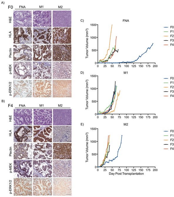
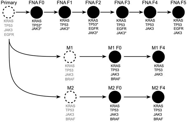
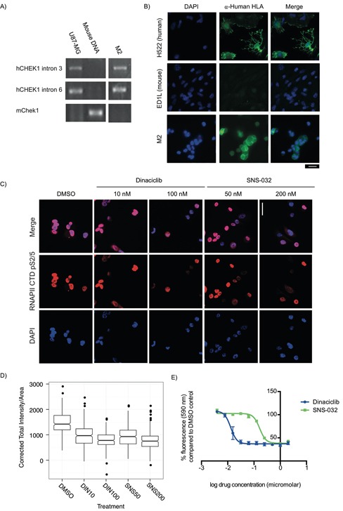
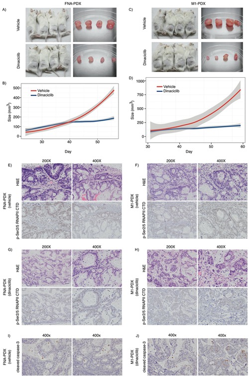
N-of-1 trials target actionable mutations, yet such approaches do not test genomically-informed therapies in patient tumor models prior to patient treatment. To address this, we developed patient-derived xenograft (PDX) models from fine needle aspiration (FNA) biopsies (FNA-PDX) obtained from primary pancreatic ductal adenocarcinoma (PDAC) at the time of diagnosis. Here, we characterize PDX models established from one primary and two metastatic sites of one patient. We identified an activating KRAS G12R mutation among other mutations in these models. In explant cells derived from these PDX tumor models with a KRAS G12R mutation, treatment with inhibitors of CDKs (including CDK9) reduced phosphorylation of a marker of CDK9 activity (phospho-RNAPII CTD Ser2/5) and reduced viability/growth of explant cells derived from PDAC PDX models. Similarly, a CDK inhibitor reduced phospho-RNAPII CTD Ser2/5, increased apoptosis, and inhibited tumor growth in FNA-PDX and patient-matched metastatic-PDX models. In summary, PDX models can be constructed from FNA biopsies of PDAC which in turn can enable genomic characterization and identification of potential therapies.

Keywords: CDK9; KRAS; fine needle aspirate biopsy; pancreatic ductal adenocarcinoma; patient-derived xenograft.


Conflict of interest statement

CONFLICTS OF INTEREST

The authors declare that they have no conflicts of interest.

Figures

Figure 1
Figure 1. Clinical pathway for establishing FNA-PDX models at time of diagnosis of pancreatic ductal adenocarcinoma
Patients enrolled in this study undergo an EUS-FNA biopsy to establish a diagnosis of PDAC prior to consideration of neoadjuvant chemotherapy and resection. During this procedure, a pooled FNA specimen is obtained for engraftment into an NSG mouse. This F0 FNA-PDX model is genomically characterized and passaged to further mice to enable evaluation of multiple therapies. Simultaneous to the development of the FNA-PDX model, patients with a diagnosis of PDAC in our cohort are enrolled onto neoadjuvant therapy protocols with subsequent resection of the tumor.
Figure 2
Figure 2. FNA-PDX and metastatic-PDX tumor growth and morphology
A-B. Representative images of H&E staining and immunohistochemical expression of human HLA (a marker specific to cells of human origin), plectin-1 (a marker of PDAC cells), phosphorylated MEK1/2 and phosphorylated ERK1/2 (markers of RAS pathway activity) in FNA-PDX and metastatic-PDX tumors for passages F0 (A) and F4 (B). Scale bars: 200 (H&E, p-ERK) μm and 100 μm (HLA plectin-1, p-MEK). C-E. FNA-PDX and patient-matched metastatic-PDX tumor morphology and growth rates of successive passaged tumors.
Figure 3
Figure 3. Next-generation sequencing of passaged FNA-PDX models revealed shared alleles with metastatic sites
Unobserved tumors (dashed circles) with inferred alleles (grey text) based on sequencing of F0-F5 primary and F0 & F4 metastatic tumors (black filled circles). Mutations identified as unique to later passage PDX models from the primary tumor are present in early passage PDX models from metastatic sites. This suggests that passaging of FNA-PDX models selects for tumor cells that match the genetic profile of the metastatic tumors. *did not meet quality thresholds for NGS and were confirmed by SNaPshot genotyping. **amplicon drop-out by NGS, confirmed by SNaPshot.
Figure 4
Figure 4. Explant cells generated from PDX models for rapid testing of targeted therapeutics are sensitive to CDK9 inhibitors
A-B. PCR primers specific to human CHEK1 and mouse Chek1 were used to identify mouse and human content in PDX explant cells, using U87-MG cells as a human positive control and mouse genomic DNA as a mouse positive control. (A) Cells from a liver metastasis explant (M2) had human CHEK1 DNA but did not have mouse Chek1 DNA. (B) To validate this, α-Human HLA class 1 A, B, and C expression (a marker of cells of human and not mouse origin) were examined by immunofluorescence. All M2 explant cells evaluated were found to be α-Human HLA A, B and/or C positive. Human (H522) and mouse (ED1L) cell lines were used as controls. Scale bar: 20 μm. C. A KRAS mutant PDX explant treated for 24 h with the CDK9 inhibitors dinaciclib (10 nM and 200 nM) and SNS-032 (50 nM and 200 nM) has reduced Ser2/5 phosphorylation of the CTD of the large subunit of RNAPII. Scale bar: 20 μm. D. Box plots of the quantification of total fluorescent intensity per unit area of cells treated with dinaciclib and SNS-032 and stained for p-Ser2/5 RNAPII CTD. Data presented was collected in three independent experiments. E. Dinaciclib (IC50: 13 nM) and SNS-032 (IC50: 165 nM) inhibit the growth of explant cells during a 3 day treatment. Data presented is the average fluorescence and corresponding standard error of the mean for three independent experiments.
Figure 5
Figure 5. Dinaciclib inhibits tumor growth in KRAS mutant FNA-PDX and metastatic-PDX models
A-D. Dinaciclib inhibits the growth of FNA-primary and patient-matched metastatic-PDX tumors in vivo. (A) FNA-PDX models established from the patient's primary tumor were treated with 40 mg/kg dinaciclib (top) or vehicle (bottom) 3 times per week for 4 weeks when the tumor reached 62.5 mm3. Tumors were excised. (B) Loess regression curve of the tumor growth of FNA-PDX models from (A) during treatment with vehicle (red) or dinaciclib (blue). 95% confidence intervals are indicated by the grey region. (C) PDX models established from a peritoneal metastasis (M1 from Figure 3) were treated with 40 mg/kg dinaciclib (top) or vehicle (bottom) and tumors excised after 4 weeks of treatment. (D) Loess regression curve from (C) during treatment with vehicle (circles) or dinaciclib (squares). 95% confidence intervals are indicated by the grey region. E-F. Representative H&E and p-RNAPII CTD Ser2/5 immunohistochemical staining of FNA-PDX tumor sections from vehicle treated mice. Tumors treated with dinaciclib exhibit reduced p-RNAPII CTD Ser2/5 as compared to the vehicle control. (G-H) Representative H&E and p-Ser2/5 RNAPII CTD immunohistochemical staining of metastatic-PDX tumor sections from dinaciclib treated mice. I-J. Representative cleaved caspase-3 immunohistochemical staining in FNA-PDX tumors from vehicle (I) and dinaciclib treated mice (J).

References

    1. Rubio-Viqueira B, Jimeno A, Cusatis G, Zhang X, Iacobuzio-Donahue C, Karikari C, Shi C, Danenberg K, Danenberg P V, Kuramochi H, Tanaka K, Singh S, Salimi-Moosavi H, et al. An in vivo platform for translational drug development in pancreatic cancer. Clin Cancer Res. 2006;12:4652–61. - PubMed
    1. Feldmann G, Mishra A, Bisht S, Karikari C, Garrido-Laguna I, Rasheed Z, Ottenhof NA, Dadon T, Alvarez H, Fendrich V, Rajeshkumar N V, Matsui W, Brossart P, et al. Cyclin-dependent kinase inhibitor Dinaciclib (SCH727965) inhibits pancreatic cancer growth and progression in murine xenograft models. Cancer Biol Ther. 2011;12:598–609. - PMC - PubMed
    1. Rubio-Viqueira B, Hidalgo M. Direct in vivo xenograft tumor model for predicting chemotherapeutic drug response in cancer patients. Clin Pharmacol Ther. 2009;85:217–21. - PubMed
    1. Kim MP, Evans DB, Wang H, Abbruzzese JL, Fleming JB, Gallick GE. Generation of orthotopic and heterotopic human pancreatic cancer xenografts in immunodeficient mice. Nat Protoc. 2009;4:1670–80. - PMC - PubMed
    1. DeRose YS, Wang G, Lin Y-C, Bernard PS, Buys SS, Ebbert MTW, Factor R, Matsen C, Milash BA, Nelson E, Neumayer L, Randall RL, Stijleman IJ, et al. Tumor grafts derived from women with breast cancer authentically reflect tumor pathology, growth, metastasis and disease outcomes. Nat Med. 2011;17:1514–20. - PMC - PubMed