Review
. 2016 Mar;10(2):166-76.
doi: 10.5009/gnl15226.

The Role of Mesothelial Cells in Liver Development, Injury, and Regeneration

Affiliations
Review

The Role of Mesothelial Cells in Liver Development, Injury, and Regeneration

Ingrid Lua et al. Gut Liver. 2016 Mar.

Abstract

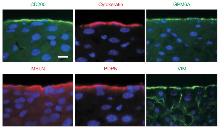
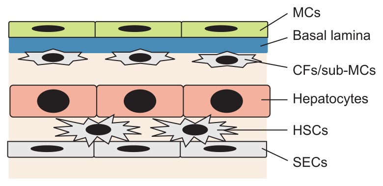
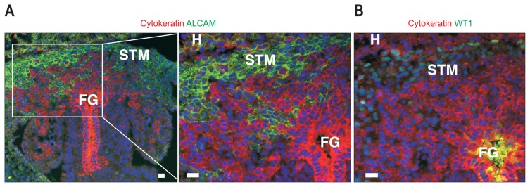
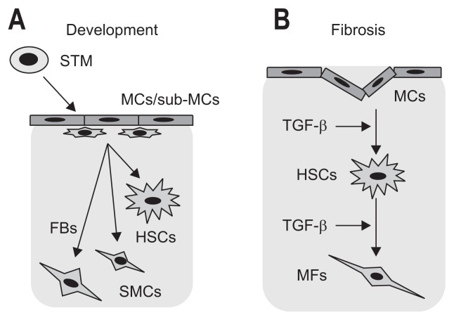
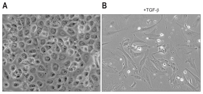
Mesothelial cells (MCs) cover the surface of visceral organs and the parietal walls of cavities, and they synthesize lubricating fluids to create a slippery surface that facilitates movement between organs without friction. Recent studies have indicated that MCs play active roles in liver development, fibrosis, and regeneration. During liver development, the mesoderm produces MCs that form a single epithelial layer of the mesothelium. MCs exhibit an intermediate phenotype between epithelial cells and mesenchymal cells. Lineage tracing studies have indicated that during liver development, MCs act as mesenchymal progenitor cells that produce hepatic stellate cells, fibroblasts around blood vessels, and smooth muscle cells. Upon liver injury, MCs migrate inward from the liver surface and produce hepatic stellate cells or myofibroblast depending on the etiology, suggesting that MCs are the source of myofibroblasts in capsular fibrosis. Similar to the activation of hepatic stellate cells, transforming growth factor β induces the conversion of MCs into myofibroblasts. Further elucidation of the biological and molecular changes involved in MC activation and fibrogenesis will contribute to the development of novel approaches for the prevention and therapy of liver fibrosis.

Keywords: Glisson's capsule; Hepatic stellate cells; Liver fibrosis; Mesothelial-mesenchymal transition; Myofibroblasts.


Figures

Fig. 1
Fig. 1
Ultrastructure of the liver surface analyzed by scanning electron microscopy. Mesothelial cells on the surface of the normal adult mouse liver have microvilli protruding into the peritoneal cavity. The black regions represent areas having few microvilli above the nuclei. Scale bars, 50 μm in (A) and 5 μm in (B).
Fig. 2
Fig. 2
Expression of markers in liver mesothelial cells (MCs). Immunohistochemistry shows the expression of CD200, cytokeratin, glycoprotein M6A (GPM6A), mesothelin (MSLN), podoplanin (PDPN), and vimentin (VIM) in MCs covering adult mouse livers. Scale bar, 10 μm.
Fig. 3
Fig. 3
Structure of the mesothelium in mouse liver tissue. Mesothelial cells (MCs) line up on the liver surface and form a single epithelial cell layer. The basal lamina separates the MCs from the underlying capsular fibroblasts (CFs)/sub-mesothelial cells (sub-MCs). Mouse livers show a single stratum of CFs beneath the MCs. Hepatic stellate cells (HSCs) reside in the space of Disse between hepatocytes and sinusoidal endothelial cells (SECs).
Fig. 4
Fig. 4
Mouse liver development at embryonic day 10. Foregut endoderm (FG) differentiates into hepatoblasts that are positive for immunohistochemical staining of cytokeratin (red). Cytokeratin+ hepatoblasts invade the surrounding septum transversum mesenchyme (STM) and form liver buds. The STM expresses mesothelial cell markers, such as activated leukocyte cell adhesion molecule (A, green) and Wilms tumor 1 homolog (WT1) (B). WT1+ mesenchymal cells also differentiate into epicardial cells of the developing heart (H). Scale bar, 20 μm.
Fig. 5
Fig. 5
Differentiation of mesothelial cells (MCs) in liver development and fibrosis. (A) During embryogenesis, mesoderm posterior 1 (MESP1)+ mesoderm gives rise to septum transversum mesenchyme (STM) and MCs. The MCs and underlying sub-MCs express Wilms tumor 1 (WT1). Cell-lineage tracing of WT1+ cells demonstrates their migration from the liver surface and differentiation into hepatic stellate cells (HSCs), fibroblasts (FBs) and smooth muscle cells (SMCs) around the blood vessels in mouse liver development. (B) Upon liver injury caused by CCl4 injection or bile duct ligation, MCs differentiate into myofibroblasts (MFs) or HSCs in adult mouse livers. Transforming growth factor β (TGF-β) provokes the mesothelial-mesenchymal transition.
Fig. 6
Fig. 6
Primary culture of liver mesothelial cells (MCs). (A) MCs isolated from adult mouse livers form epithelial colonies in culture. (B) Transforming growth factor β (TGF-β) treatment induces the mesothelial-mesenchymal transition.

References

    1. Minot CS. The mesoderm and the coelom of vertebrates. Am Nat. 1890;24:877–898. doi: 10.1086/275198. - DOI
    1. Mutsaers SE. Mesothelial cells: their structure, function and role in serosal repair. Respirology. 2002;7:171–191. doi: 10.1046/j.1440-1843.2002.00404.x. - DOI - PubMed
    1. Jones JS. Pathology of the mesothelium. London: Springer-Verlag; 1987. - DOI
    1. Simionescu M, Simionescu N. Organization of cell junctions in the peritoneal mesothelium. J Cell Biol. 1977;74:98–110. doi: 10.1083/jcb.74.1.98. - DOI - PMC - PubMed
    1. Yung S, Thomas GJ, Stylianou E, Williams JD, Coles GA, Davies M. Source of peritoneal proteoglycans: human peritoneal mesothelial cells synthesize and secrete mainly small dermatan sulfate proteoglycans. Am J Pathol. 1995;146:520–529. - PMC - PubMed

Publication types

LinkOut - more resources