Review
. 2016 Apr;17(4):224-38.
doi: 10.1038/nrn.2016.9. Epub 2016 Mar 3.

Decoding the organization of spinal circuits that control locomotion

Affiliations
Review

Decoding the organization of spinal circuits that control locomotion

Ole Kiehn. Nat Rev Neurosci. 2016 Apr.

Abstract

Unravelling the functional operation of neuronal networks and linking cellular activity to specific behavioural outcomes are among the biggest challenges in neuroscience. In this broad field of research, substantial progress has been made in studies of the spinal networks that control locomotion. Through united efforts using electrophysiological and molecular genetic network approaches and behavioural studies in phylogenetically diverse experimental models, the organization of locomotor networks has begun to be decoded. The emergent themes from this research are that the locomotor networks have a modular organization with distinct transmitter and molecular codes and that their organization is reconfigured with changes to the speed of locomotion or changes in gait.


Figures

Figure 1
Figure 1. Organization of neuronal control of locomotion in vertebrates
The selection and initiation of locomotor behaviour involves various regions of the brain and brainstem. Output neurons of the basal ganglia (BG) project both to the thalamus (Tha) — which sends projections to the motor cortex (MCtx) and other cortical areas — and to areas in the brainstem, including the mesencephalic locomotor region (MLR). Initiation of locomotion is thought to be mediated by the activity of neurons in the MLR,, including the cuneiform nucleus (CNf) and the pedunculopontine nucleus (PPN). MLR neurons project to neurons in the reticular formation (RF) in the hindbrain,. Neurons in the RF project to locomotor networks in the spinal cord that execute locomotion. Descending fibres from the vestibular and rubrospinal spinal pathways (brainstem nuclei (BSN)) maintain posture and modulatory signals,– that regulate the ongoing locomotor activity. The cerebellum coordinates locomotor behaviour by mediating movement-generated feedback and internal feedback, as well as by modulating the activity in the descending pathways. Proprioceptive sensory feedback modulates the activity of the spinal locomotor network. Cortical activity (MCtx) provides visuomotor (VCtx) correction of locomotion via the posterior parietal cortex (pPCtx). Figure is adapted with permission from REF. 165, Springer.
Figure 2
Figure 2. Multiple left–right coordination circuits
Diverse commissural neurons (CNs) control left–right coordination at different frequencies of locomotion in mice. a | V0 CNs develop from the p0 progenitor domain in the ventral spinal cord. There are five specific progenitor domains (p0–p3 and pMN) in the ventral cord and six in the dorsal cord (pd1–pd6), each of which is characterized by differential expression of transcription factors (see REFS 29,84,166). When the progenitor cells mature, they migrate laterally and become neurons or motor neurons. p0 progenitors express developing brain homeobox 1 (DBX1) and develop into inhibitory dorsal V0 neurons (V0D neurons), which express paired box protein 7 PAX7 (PAX7+) but not homeobox even-skipped homologue protein 1 (EVX1) (EVX1), and excitatory ventral V0 neurons (V0V neurons), which are PAX7 but EVX1+. b | Genetic ablation of V0 neurons leads to the loss of hindlimb alternation at all locomotor frequencies in vitro, as indicated by the switch from out-of-phase left–right locomotor-like activity in wild-type mice (two upper rows) to in-phase locomotor-like activity (third and fourth rows). V0D neurons secure alternation at low frequencies: deletion of V0D neurons leads to synchronous left–right locomotor-like activity at low frequencies but maintained alternation at high frequencies of locomotor-like activity (fifth and sixth rows). V0V neurons secure hindlimb alternation at high frequencies of locomotion: deletion of the V0V neurons leads to synchronous left–right activity at high frequencies but maintained alternation at low frequencies of locomotor-like activity (seventh and eight rows). c | In the presence of V0 neurons, mice express four gaits at different frequencies of locomotion: walk, trot, gallop and bound. When V0 neurons are ablated, mice only express bound at all frequencies of locomotion, whereas when V0V neurons are ablated, mice lack trot but can express walk and gallop, which is now expressed in the frequency range at which trot is normally expressed. V0D neuron-ablated animals do not survive postnatally and therefore their gaits were not tested. d | Proposed recruitment of V0D- and V0V-related pathways in response to increased locomotor frequency. At low frequencies of locomotion that correspond to walk (left-hand panel), inhibitory V0D CNs are activated by rhythm-generating neurons (R) on the same side of the cord. Their activation leads to the inhibition of locomotor networks on the other side of the cord, including motor neurons (MNs). At higher frequencies of locomotion that correspond to trot excitatory (middle panel), V0V commissural neurons are recruited. Their activation causes the inhibition of locomotor networks on the other side of the cord, including MNs via local inhibitory neurons (blue). At very high frequencies of locomotion that correspond to bound (right-hand panel) left–right synchrony is secured by excitatory non-V0 neurons (red), which are possibly V3 neurons that originate from single-minded homologue 1 (SIM1)-expressing progenitor cells. Note that a single neuron in the diagrams represents a group of neurons. e | Proposed CN network in lamprey and tadpole. The core of the network is made up of inhibitory CNs that cross the midline and inhibit excitatory neurons rhythm-generating neurons and MNs on the other side of the cord (as indicated by the square box). Part a is based on data from REFS 48–. Part b is based on data from REF. 50. Part c is based on data from REF. 51. Part d is based on data from REFS 47,,,,. Part e is based on data presented in REFS 11,,,.
Figure 3
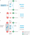
Figure 3. Organizational and molecular delineation of rhythm-generating circuits
Rhythm-generating circuits are excitatory in all the vertebrates that have been investigated. a,b | In tadpoles, the circuit is composed of reciprocally and electrically connected glutamatergic and cholinergic neurons (excitatory interneurons (EINs)) that are located in the hindbrain and the spinal cord. In lampreys, EINs are glutamatergic, with synaptic connections to other EINs, and are located in each segment along the spinal cord. EINs drive motor neurons (MNs; which project to the muscles) and inhibitory commissural neurons (CNs; with axons projecting to the other half of the cord) on the same side of the cord. Inhibitory neurons with connections on the same side of the cord are omitted in the diagrams (for example, see REF. 26). c | Proposed circuits for the rhythm-generating circuit in the mouse spinal cord are shown. The rhythm-generating circuit (R) is composed of neurons that express the transcription factor short stature homeobox protein 2 (SHOX2). Rhythm-generating circuits drive left–right alternating circuits (V0D–V0V), including V2a neurons that express the transcription factor ceh 10 homeodomain containing homologue (CHX10), and neurons that are both CHX10- and SHOX2-positive (V2a SHOX2+) that presumably connect to motor neurons. Rhythm-generation circuits also drive left–right synchronizing circuits (non-V0, possibly of the V3 class). Only the left side of the circuit is shown. Blocking the synaptic output of SHOX2+ neurons or optogenetic silencing these neurons disrupts the rhythm without completely abolishing it, suggesting that as yet unidentified EINs contribute to the rhythm. d | The rhythm-generating circuits in zebrafish larvae are composed of CNs that belong to the excitatory multipolar commissural descending type (MCoD) neurons and circumferential ipsilateral descending (CID) neurons, which are analogues of V0V and V2a mouse neurons, respectively. The MCoDs (CNs) are active at low swimming frequencies but are silenced as the swimming frequencies increase (>40 Hz). The probability of CID (V2a) neuron firing increases with frequency, with the most dorsal neuron active at the highest frequencies (60–90 Hz). Laser ablation of MCoD neurons abrogates slow swimming, whereas ablation of dorsal CID (V2a) neurons abolishes high frequency swimming frequency. e | In the adult zebrafish, three groups of rhythm-generating V2a neurons innervate slow, intermediate and fast MNs. The three groups of V2a neurons are recruited incrementally (as indicated by the colour change) in a modular manner that reflects an ordered recruitment of slow, intermediate and fast MNs as the speed of swimming increases. Parts a and b are based on data from REFS 11,,,,. Part c is based on data from REFS 50,,,. Part d is based on data from REFS 30,,,,. Part e is based on data from REFS 25,.
Figure 4
Figure 4. Multiple levels of flexor–extensor antagonism
During limbed locomotion, motor neurons (MNs) that control flexor and extensor muscles around different joints within a limb must be activated in alternation. This flexor and extensor antagonism is generated by the activity of inhibitory neurons that are active at multiple levels in the locomotor network. a | One synapse away from flexor and extensor MNs are reciprocal Ia-inhibitory neurons (rIa-INs), which reciprocally inhibit antagonist MNs and each other. A minimal network of rIa-INs may coordinate flexor–extensor alternation (out of phase; denoted by blue boxes) in the isolated spinal cord in the absence of excitatory rhythm-generating circuits when appropriately activated by drugs. b | When two major groups of inhibitory neurons in the ventral spinal cord, V1 and V2b inhibitory neurons, are genetically ablated, all flexor–extensor alternation is lost leaving only flexor–extensor synchrony during locomotor-like activity (in phase; blue boxes). Excitatory neurons of different kinds provide premotor rhythmic excitation. `R' refers to rhythm-generating neurons. c | Schematic showing multiple levels of control of flexor–extensor alternation with all circuit elements intact. A module comprising excitatory neurons and rIa-INs (dashed boxes) receives input from excitatory rhythm-generating circuits and provides rhythmic excitation and inhibition to flexor and extensor antagonism, respectively. Inhibitory neurons belonging to the V1 and V2b classes of neurons (blue box) provide reciprocal inhibition between flexor and extensor rhythm generators. rIa-Ins also belong to the V1 and V2b classes. Part a is based on data from REF. 123. Part b is based on data from REF. 134. Part c is based on data from REFS 119,,.
Figure 5
Figure 5. Proprioceptive input directs stance and swing phase transitions
The activity of the locomotor network is regulated by sensory signals such as proprioceptive sensory information that is generated by active movement. a | Proprioceptors in hip muscles are activated when the hip is flexed and extended. When the hip is fully extended (step 1) there is maximal stretch-mediated proprioception from flexor hip muscles, which increases the excitation of the flexor (F) rhythm generator. Unloading of the ankle extensor Golgi tendon organs (GTOs) at the end of the stance phase (step 2) decreases the drive to the extensor (E) rhythm generator. The hip and ankle proprioceptors therefore act in synergy on the locomotor network to increase excitation and to decrease inhibition of the flexor rhythm generator at the end of the stance phase, promoting swing-to-stance phase transition. bd | Genetic elimination of stretch-activated (from muscle spindles) and force-activated (from GTOs) proprioception leads to changes in locomotor patterns in mice. The green and blue bars indicate the times of activation for hip, knee and ankle flexors and extensors with respect to the swing phase (light grey) and stance phase (light blue). Elimination of muscle spindle afferents prolongs ankle flexor swing-to-stance phase transition, whereas elimination of both the muscle spindles and the GTOs leads to phase advance of knee and ankle flexors with respect to the hip flexor, corresponding to a removal of the indirect inhibition of the flexor rhythm generator from GTOs. Part a based on data from REFS 17,–. Parts bd are based on data from REFS 144,.

References

    1. Drew T, Marigold DS. Taking the next step: cortical contributions to the control of locomotion. Curr. Opin. Neurobiol. 2015;33:25–33. - PubMed
    1. Takakusaki K. Neurophysiology of gait: from the spinal cord to the frontal lobe. Mov. Disord. 2013;28:1483–1491. - PubMed
    1. Garcia-Rill E. The basal ganglia and the locomotor regions. Brain Res. 1986;396:47–63. - PubMed
    1. Grillner S, Robertson B. The basal ganglia downstream control of brainstem motor centres — an evolutionarily conserved strategy. Curr. Opin. Neurobiol. 2015;33:47–52. - PubMed
    1. Garcia-Rill E, Hyde J, Kezunovic N, Urbano FJ, Petersen E. The physiology of the pedunculopontine nucleus: implications for deep brain stimulation. J. Neural Transmission. 2015;122:225–235. - PMC - PubMed

Publication types