. 2016 Apr;37(4):958-66.
doi: 10.3892/ijmm.2016.2488. Epub 2016 Feb 12.

Role of Alix in miRNA packaging during extracellular vesicle biogenesis

Affiliations

Role of Alix in miRNA packaging during extracellular vesicle biogenesis

Alessandra Iavello et al. Int J Mol Med. 2016 Apr.

Abstract

Evidence indicates that Alix, an accessory protein of the endosomal sorting complex required for transport (ESCRT), is involved in the biogenesis of extracellular vesicles (EVs). EVs contain selected patterns of microRNAs (miRNAs or miRs); however, little is known about the mechanisms of miRNA enrichment in EVs. The aim of the present study was to evaluate whether Alix is involved in the packaging of miRNAs within EVs released by human liver stem‑like cells (HLSCs). EVs released from HLSCs were enriched with miRNAs and expressed Alix and several RNA-binding proteins, including Argonaute 2 (Ago2), a member of the Argonaute family known to be involved in the transport and the processing of miRNAs. Co-immunoprecipitation experiments revealed an association between Alix and Ago2. The results from RT-qPCR indicated that in the Alix/Ago2 immunoprecipitates, miRNAs were detectable. EVs were instrumental in transferring selected miRNAs from HLSCs to human endothelial cells absent in the latter cells. Alix knockdown did not influence the number of EVs released by HLSCs, but it significantly decreased miRNA expression levels in the EVs and consequently their transfer to the endothelium. Our findings indicate that Alix binds to Ago2 and miRNAs, suggesting that it plays a key role in miRNA enrichment during EV biogenesis. These results may represent a novel function of Alix, demonstrating its involvement in the EV-mediated transfer of miRNAs.


Figures

Figure 1
Figure 1
Characterization of human liver stem-like cell (HLSC)-derived extracellular vesicles (EVs; HLSC-EVs). (A) Representative NanoSight image of 100k g purified EVs; inset representative transmission electron microscopy image of purified EVs negatively stained with NanoVan (black line=100 nm). (B) Representative western blots of CD63 expression in HLSC-derived EVs. (C) Representative western blots of endosomal sorting complex required for transport (ESCRT) components (Tsg101, CHMP4 a,b,c, Alix and STAM) performed on HLSCs and HLSC-derived EVs. (D) Representative western blots showing the expression of ribonucleoproteins (TIA, TIAR, HuR, Stau1/2 and Ago2) and RPS29 in HLSCs and HLSC-derived EVs. Four experiments were performed with similar results. (E) Representative immunogold electron microscopy images showing staining for Ago2 (upper panel) and control where the primary antibody was omitted (lower panel). EVs were observed using a JEOL Jem-1010 electron microscope (black line, 100 nm).
Figure 2
Figure 2
Alix and Ago2 co-immunoprecipitation (Co-IP). (A) Representative western blots and densitometric analysis of Alix and Ago2 expression in human liver stem-like cell (HLSC)-derived extracellular vesicles (EVs; HLSC-EVs) at 3, 6, 16, 24 h of cell starvation. (B) Representative Co-IP experiments performed on HLSCs (top panel) and HLSC-derived EVs (bottom panel) revealed interaction between Alix and Ago2. The immune complexes were formed by pre-incubation with anti-Ago2 (IP Ago2) and revealed with Alix antibody (left panels), or by pre-incubation with anti-Alix (IP Alix panels) and revealed with Ago2 antibody (right). For IP-positive control (CTR+), to evaluate the effective immunoprecipitation, the membranes were blotted with an antibody that recognises the same immunoprecipitated antigen. As a negative control (CTR−) non-immune IgG were used. Six experiments were performed with similar results.
Figure 3
Figure 3
miRNA expression in human liver stem-like cell (HLSC)-derived extracellular vesicles (EVs; HLSC-EVs). (A) The expression of miR24, miR31, miR125b, miR99b, miR221, miR16 and miR451 was evaluated by RT-qPCR in HLSC-derived EVs with respect to HLSCs. Data are expressed as log of relative quantification (Rq), normalized to RNU6B and to one for HLSC. These miRNAs were enriched in EVs in respect to their relative HLSC. Data are the means ± SD of 4 experiments. (B) The expression of miR24, miR31, miR125b, miR99b, miR221, miR16 and miR451 was evaluated by RT-qPCR in HLSC-derived EVs immunoprecipitated with Ago2 (gray bars) and in HLSC-derived EVs immunoprecipitated with Alix (black bars) with respect to 100k g purified EVs. Data are expressed as the log of Rq, normalized to RNU6B and to one for 100k g purified EVs. Data are the means ± SD of 4 experiments.
Figure 4
Figure 4
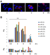
Extracellular vesicle (EV) internalization and miRNA transfer in human umbilical cord vein-derived endothelial cells (HUVECs). (A) Representative image of internalization of PKH26-labeled human liver stem-like cell-derived EVs into HUVECs detected by confocal microscopy at different time-points. Three experiments were performed with similar results. (B, upper panel) RT-qPCR performed on HUVEC RNA treated with actinomycin D (ActD), in order to block RNA transcription, and incubated with HLSC-derived EVs at different time-points (3, 6 and 12 h). HUVECs without EV stimulation at time 0 were used as control (Ctr) and set to one. (B lower panel) RT-qPCR performed on HUVEC RNA treated with ActD, in order to block RNA transcription, and incubated with vehicle alone at different times (3, 6 and 12 h). Data are expressed as relative quantification (Rq), normalized to RNU6B. Data are the means ± SD of 4 experiments. *P<0.05; **P<0.001 vs. control (Ctr).
Figure 5
Figure 5
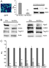
Effects of Alix knockdown in human liver stem-like cells (HLSCs). (A) Representative image of HLSCs transfected with the positive control vector, pmaxGFP, 24 h after transfection (original magnification, ×100); Alix mRNA expression in HLSCs in which Alix was knocked down (kd-HLSCs) with respect to normal HLSCs (Ctr HLSCs) set to one, was evaluated by RT-qPCR. Data are the means ± SD of 3 experiments expressed as relative quantification (Rq), normalized to 18S. Representative western blots of Alix protein expression in kd-HLSCs with respect to Ctr HLSCs. Equal loading was confirmed by stripping the immunoblot and reprobing it for actin. Three different experiments were performed with similar results. (B) Representative western blots of Alix and Ago2 protein expression in extracellular vesicles (EVs) released from normal HLSCs (EVs) and in EVs released from HLSCs in which Alix was knocked down (kd-EVs). Equal loading was confirmed by stripping the immunoblot and reprobing it for CD63 and Tsg101. Three different experiments were performed with similar results. (C) The expression of miR-24, miR-31, miR-125b, miR-99b, miR-221, miR-16 and miR-451 was evaluated by RT-qPCR in EVs derived from HLSCs in which Alix was knocked down (gray bars) with respect to EVs derived from normal HLSCs (black bars) set to one. Data are expressed as Rq, normalized to RNU6B. Data are the means ± SD of 4 experiments. **P<0.001 vs. HLSC-derived EVs.
Figure 6
Figure 6
Effect of Alix knockdown on extracellular vesicle (EV)-mediated transfer of miRNAs to human umbilical cord vein-derived endothelial cells (HUVECs). RT-qPCR performed on RNA extracted from HUVECs treated with actinomycin D (ActD) and incubated with human liver stem-like cell (HLSC)-derived EVs (EV) or EVs derived from HLSCs in which Alix was knocked down (kd-EVs) for 3 h. HUVECs without EV stimulation were used as controls (Ctr) and set to one. Data are expressed as relative quantification (Rq), normalized to RNU6B. Data are the means ± SD of 4 experiments. **P<0.001 kd-EVs vs. EVs.

References

    1. Quesenberry PJ, Goldberg LR, Aliotta JM, Dooner MS, Pereira MG, Wen S, Camussi G. Cellular phenotype and extracellular vesicles: basic and clinical considerations. Stem Cells Dev. 2014;23:1429–1436. doi: 10.1089/scd.2013.0594. - DOI - PMC - PubMed
    1. Ratajczak J, Wysoczynski M, Hayek F, Janowska-Wieczorek A, Ratajczak MZ. Membrane-derived microvesicles: important and underappreciated mediators of cell-to-cell communication. Leukemia. 2006;20:1487–1495. doi: 10.1038/sj.leu.2404296. - DOI - PubMed
    1. Théry C, Ostrowski M, Segura E. Membrane vesicles as conveyors of immune responses. Nat Rev Immunol. 2009;9:581–593. doi: 10.1038/nri2567. - DOI - PubMed
    1. Lee TH, D'Asti E, Magnus N, Al-Nedawi K, Meehan B, Rak J. Microvesicles as mediators of intercellular communication in cancer - the emerging science of cellular 'debris'. Semin Immunopathol. 2011;33:455–467. doi: 10.1007/s00281-011-0250-3. - DOI - PubMed
    1. Cocucci E, Racchetti G, Meldolesi J. Shedding microvesicles: artefacts no more. Trends Cell Biol. 2009;19:43–51. doi: 10.1016/j.tcb.2008.11.003. - DOI - PubMed

Publication types

MeSH terms