. 2016 May;9(5):406-414.
doi: 10.1158/1940-6207.CAPR-15-0347. Epub 2016 Mar 2.

Inflammation-Related IL1β/IL1R Signaling Promotes the Development of Asbestos-Induced Malignant Mesothelioma

Affiliations

Inflammation-Related IL1β/IL1R Signaling Promotes the Development of Asbestos-Induced Malignant Mesothelioma

Yuwaraj Kadariya et al. Cancer Prev Res (Phila). 2016 May.

Abstract

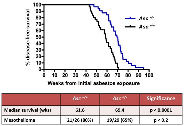
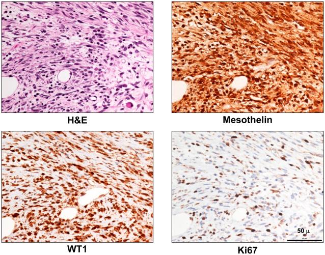
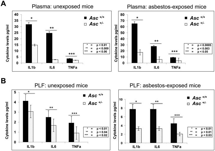
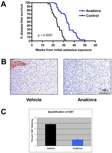
Exposure to asbestos is causally associated with the development of malignant mesothelioma, a cancer of cells lining the internal body cavities. Malignant mesothelioma is an aggressive cancer resistant to all current therapies. Once inhaled or ingested, asbestos causes inflammation in and around tissues that come in contact with these carcinogenic fibers. Recent studies suggest that inflammation is a major contributing factor in the development of many types of cancer, including malignant mesothelioma. The NALP3/NLRP3 inflammasome, including the component ASC, is thought to be an important mediator of inflammation in cells that sense extracellular insults, such as asbestos, and activate a signaling cascade resulting in release of mature IL1β and recruitment of inflammatory cells. To determine if inflammasome-mediated inflammation contributes to asbestos-induced malignant mesothelioma, we chronically exposed Asc-deficient mice and wild-type littermates to asbestos and evaluated differences in tumor incidence and latency. The Asc-deficient mice showed significantly delayed tumor onset and reduced malignant mesothelioma incidence compared with wild-type animals. We also tested whether inflammation-related release of IL1β contributes to tumor development in an accelerated mouse model of asbestos-induced malignant mesothelioma. Nf2(+/-);Cdkn2a(+/-) mice exposed to asbestos in the presence of anakinra, an IL1 receptor (IL1R) antagonist, showed a marked delay in the median time of malignant mesothelioma onset compared with similarly exposed mice given vehicle control (33.1 weeks vs. 22.6 weeks, respectively). Collectively, these studies provide evidence for a link between inflammation-related IL1β/IL1R signaling and the development of asbestos-induced malignant mesothelioma. Furthermore, these findings provide rationale for chemoprevention strategies targeting IL1β/IL1R signaling in high-risk, asbestos-exposed populations. Cancer Prev Res; 9(5); 406-14. ©2016 AACR.


Figures

Figure 1
Figure 1
Asc-deficiency significantly delays malignant mesothelioma (MM) development in mice chronically exposed to asbestos. Top, Kaplan-Meier curves of percent disease-free animals demonstrate increased latency of asbestos-induced MM in heterozygous Asc (Asc+/− ) mice than in asbestos-treated wild-type (WT, Asc +/+) littermates. Bottom, summary of differences in median percent disease-free animals and MM incidence in Asc+/− and WT mice. Note that the difference in the percentage of disease-free animals, but not MM incidence, between WT and Asc+/− mice was highly significant (p < 0.0001). However, the difference in MM incidence between WT and homozygous Asc−/− mice was statistically significant (p < 0.05) (see Supplemental Figs. S1 and S2).
Figure 2
Figure 2
Histopathologic assessment of MM in an Asc+/− mouse. Serial tumor sections depicting H&E staining (top left panel) and IHC for MM markers mesothelin (top right) and WT1 (bottom left) in a representative MM from an Asc+/− mouse; Ki67 staining (lower right) indicates active cellular proliferation in the MM cells. All original images are at 400X magnification. Scale bar = 50 μ.
Figure 3
Figure 3
Decreased levels of IL-1β and other inflammatory cytokines in serum and peritoneal lavage fluid (PLF) from Asc+/− mice compared to that of WT (Asc+/+) mice. A) IL-1β, IL-6 and TNFα levels in serum of Asc+/− and Asc +/+ mice exposed or not (unexposed) to asbestos. B) IL-1β, IL-6 and TNFα levels in PLF of Asc+/− and Asc +/+ mice exposed or unexposed to asbestos. P-values for differences between the indicated samples from Asc+/+ (black bars) and Asc+/− (white bars) mice are provided for each cytokine and experimental condition tested.
Figure 4
Figure 4
Anakinra treatment delays tumor onset and inhibits cell proliferation in a mouse model of accelerated asbestos-induced MM. A) Kaplan-Meier curves of asbestos-exposed Nf2+/−;Cdkn2a+/− mice treated with anakinra or vehicle (citrate buffer). Treatment of asbestos-exposed Nf2+/−;Cdkn2a+/− mice with anakinra resulted in significantly prolonged survival compared to that observed in vehicle-treated littermates. B) Ki67 nuclear staining (brown) of MMs from vehicle- and anakinra-treated mice. Note that adjacent normal liver in vehicle-treated mouse (upper left) shows non-specific cytoplasmic staining. Original images: 200X magnification. Representative 100-μm scale bar is shown. C) Quantification of the average percentage of Ki67-positive nuclei in MM tumors from mice treated with anakinra or vehicle. Error bars represent standard deviation of multiple tumor sections from 6-7 mice per arm.
Figure 5
Figure 5
Anakinra inhibits IL-1β/IL-1R signaling both in vitro and in vivo. A) Immunoblot analysis of normal human mesothelial cells (HM3) starved overnight and then stimulated with IL-1β in the presence or absence of anakinra. Anakinra-treated cells show markedly decreased activity of AKT, ERK and JNK, as illustrated by decreased expression using phospho (P)-specific antibodies. B) Immunohistochemical staining (reddish brown) for P-AKT, P-JNK and P-ERK in MMs derived from asbestos-exposed mice treated or not with anakinra. Original images: 200X magnification. Scale bar = 100 μ.

References

    1. Wagner JC, Slegga CA, Marchand P. Diffuse pleural mesothelioma and asbestos exposure in the North Western Cape Province. Br J Ind Med. 1960;17:260–71. - PMC - PubMed
    1. Henley SJ, Larson TC, Wu M, Antao VC, Lewis M, Pinheiro GA, et al. Mesothelioma incidence in 50 states and the District of Columbia, United States, 2003-2008. Int J Occup Environ Health. 2013;19:1–10. - PMC - PubMed
    1. Goldberg M, Imbernon E, Rolland P, Gilg Soit Ilg A, Savès M, de Quillacq A, et al. The French National Mesothelioma Surveillance Program. Occup Environ Med. 2006;63:390–5. - PMC - PubMed
    1. Burki T. Health experts concerned over India's asbestos industry. Lancet. 2010;375:626–7. - PubMed
    1. Flejter WL, Li FP, Antman KH, Testa JR. Recurring loss involving chromosomes 1, 3, and 22 in malignant mesothelioma: possible sites of tumor suppressor genes. Genes, Chromosomes Cancer. 1989;1:148–54. - PubMed

Publication types

MeSH terms