. 2016 Mar 3;531(7592):118-21.
doi: 10.1038/nature17200.

Pre-fusion structure of a human coronavirus spike protein

Affiliations

Pre-fusion structure of a human coronavirus spike protein

Robert N Kirchdoerfer et al. Nature. .

Abstract

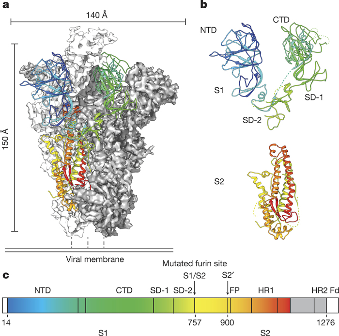
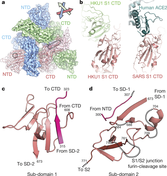
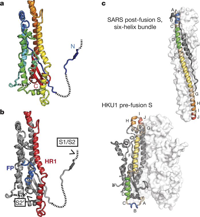
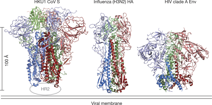
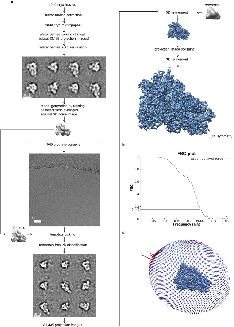
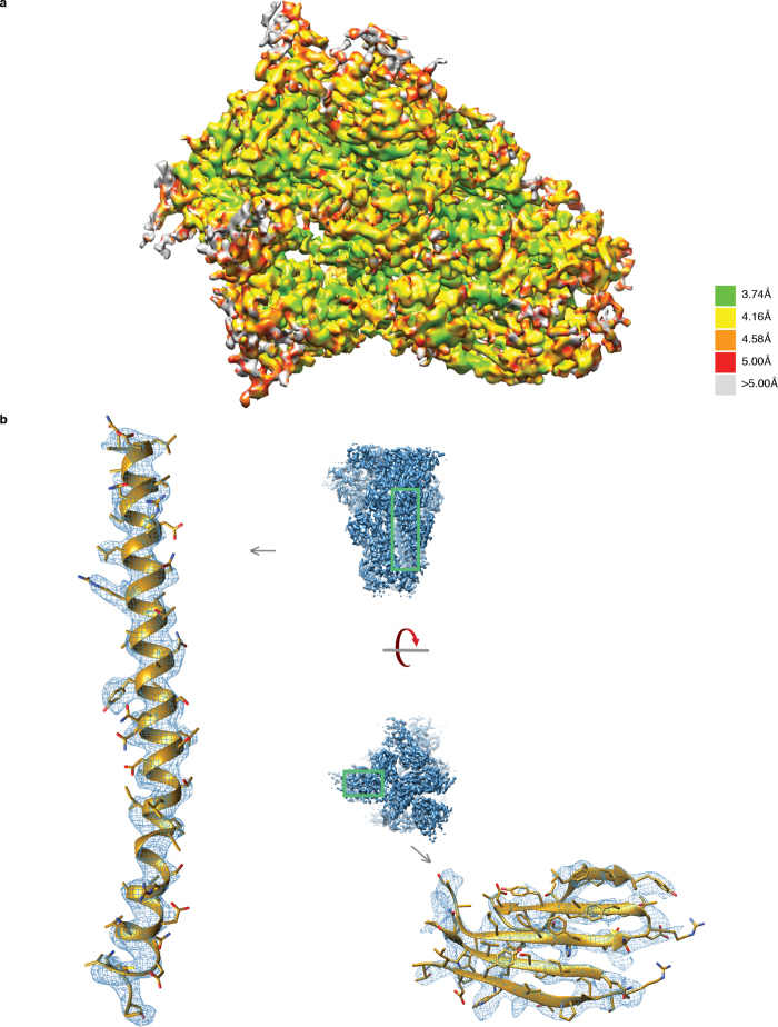
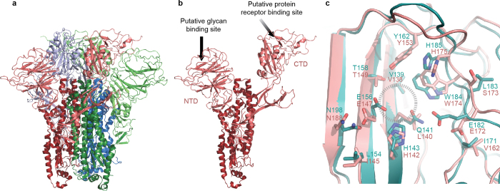
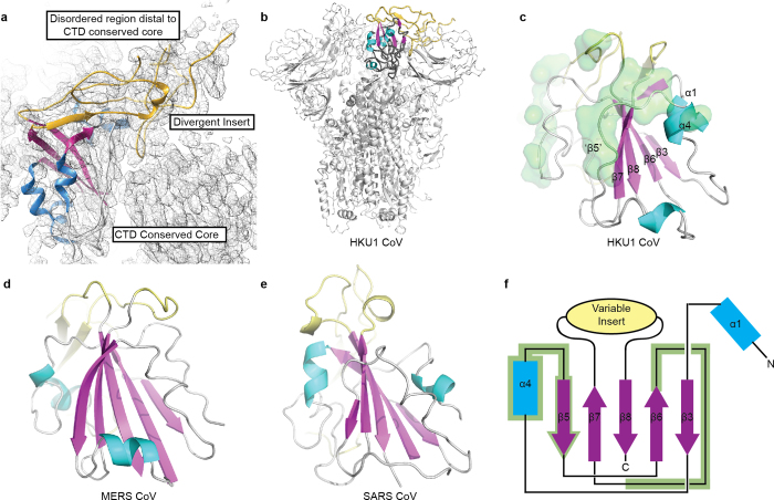
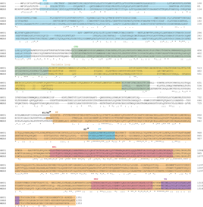
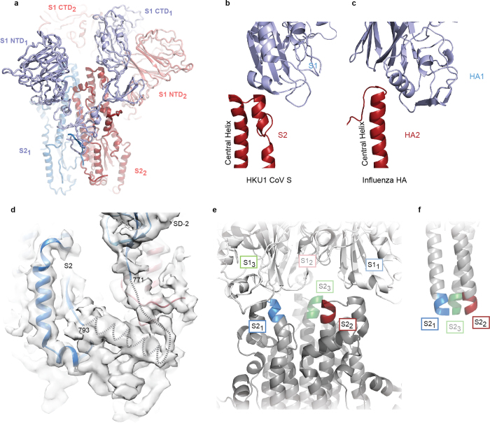
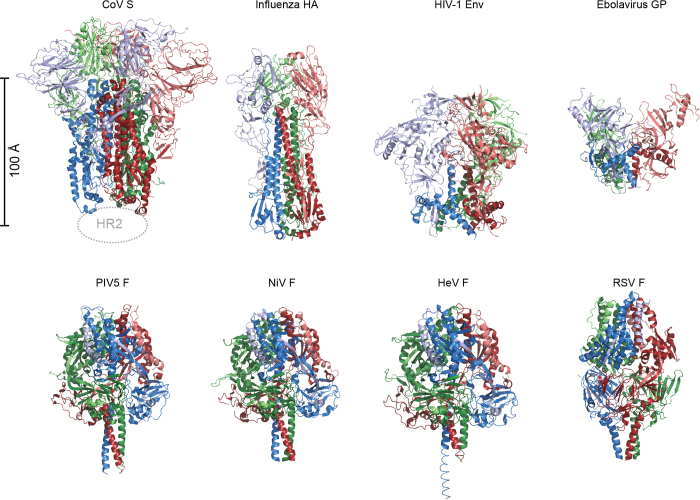
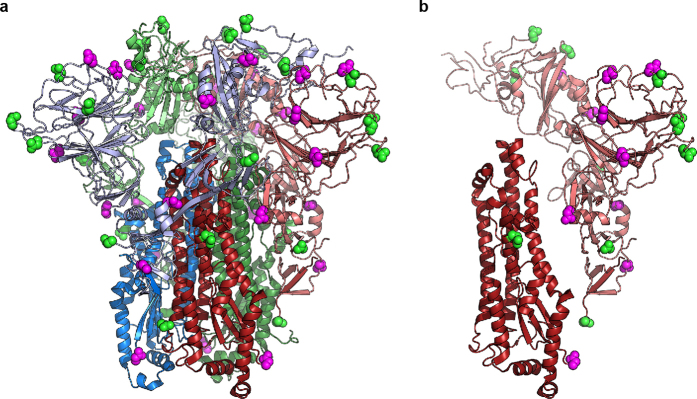
HKU1 is a human betacoronavirus that causes mild yet prevalent respiratory disease, and is related to the zoonotic SARS and MERS betacoronaviruses, which have high fatality rates and pandemic potential. Cell tropism and host range is determined in part by the coronavirus spike (S) protein, which binds cellular receptors and mediates membrane fusion. As the largest known class I fusion protein, its size and extensive glycosylation have hindered structural studies of the full ectodomain, thus preventing a molecular understanding of its function and limiting development of effective interventions. Here we present the 4.0 Å resolution structure of the trimeric HKU1 S protein determined using single-particle cryo-electron microscopy. In the pre-fusion conformation, the receptor-binding subunits, S1, rest above the fusion-mediating subunits, S2, preventing their conformational rearrangement. Surprisingly, the S1 C-terminal domains are interdigitated and form extensive quaternary interactions that occlude surfaces known in other coronaviruses to bind protein receptors. These features, along with the location of the two protease sites known to be important for coronavirus entry, provide a structural basis to support a model of membrane fusion mediated by progressive S protein destabilization through receptor binding and proteolytic cleavage. These studies should also serve as a foundation for the structure-based design of betacoronavirus vaccine immunogens.


Conflict of interest statement

The authors declare no competing financial interests.

Figures

Figure 1
Figure 1. Structure of the HKU1 pre-fusion spike ectodomain.
a, A single protomer of the trimeric S protein is shown in cartoon representation coloured as a rainbow from the N to C terminus (blue to red) with the reconstructed EM density of remaining protomers shown in white and grey. b, The S1 subunit is composed of the NTD and CTD as well as two sub-domains (SD-1 and SD-2). The S2 subunit contains the coronavirus fusion machinery and is primarily α-helical. c, Domain architecture of the HKU1 S protein coloured as in a. PowerPoint slide
Figure 2
Figure 2. Architecture of the HKU1 S1 subunit.
a, EM density corresponding to each S1 protomer is shown. The putative glycan-binding and protein-receptor-binding sites are indicated with dashed shapes on the NTD and CTD, respectively. b, The HKU1 S1 CTD forms quaternary interactions with an adjacent CTD using a surface similar to that used by SARS CTD to bind its receptor, ACE2 (ref. 11). c, Sub-domain 1 is composed of amino acid residues before and after the S1 CTD. d, Sub-domain 2 is composed of S1 sequence C-terminal to the CTD, a short peptide following the NTD, and the N-terminal strand of S2, which follows the S1/S2 furin-cleavage site. PowerPoint slide
Figure 3
Figure 3. HKU1 S2 subunit fusion machinery.
a, The HKU1 S2 subunit is coloured like a rainbow from the N-terminal β-strand (blue), which participates in S1 sub-domain 2, to the C terminus (red) before HR2. b, The HKU1 S2 structure contains the fusion peptide (FP) and a heptad repeat (HR1). Protease-recognition sites are indicated within disordered regions of the protein (dashed lines). c, A comparison of coronavirus S2 HR1 in the pre- and post-fusion conformations. Five HR1 α-helices are labelled and coloured like a rainbow from blue to red, N to C terminus, respectively. The structures are oriented to position similar portions of the central helix (red). PowerPoint slide
Figure 4
Figure 4. Comparison of structurally related class I viral fusion proteins.
The fusion proteins from coronaviruses, influenza virus and HIV-1 are cleaved into receptor-binding subunits (pink, light green, light blue) and the viral fusion machinery (dark red, dark green, blue),,,. Comparison to other class I fusion proteins can be found in Extended Data Fig. 8. PowerPoint slide
Extended Data Figure 1
Extended Data Figure 1. Data processing flowchart.
a, Processing resulting in density map of pre-fusion HKU1 spike glycoprotein at 4.04 Å resolution. b, FSC plot illustrating correlation between two volumes refined independently from two distinct half sets of raw data. A final resolution of 4.04 Å is indicated in the plot. c, Angular distribution of raw data within the data set. A slight, but within normal range, over-representation of top views was observed (tall red bars).
Extended Data Figure 2
Extended Data Figure 2. Resolution of the pre-fusion HKU1 S density map.
a, Local resolution within the EM density map. Local resolution was calculated using ResMap discretizing every 0.25 Å over a range from 2 × voxel size (2.62 Å) to 4 × voxel size (5.24 Å). Resolution significance criterion was set to 0.05. The resolution ranges from 3.74 Å in stable internal secondary structures to greater than 5.00 Å in flexible peripheral loops. b, Close-ups of secondary-structure densities. To the left is displayed the central α-helix of an S2 monomer and to the right is a β-sheet from the NTD domain in an S1 monomer.
Extended Data Figure 3
Extended Data Figure 3. Cleavage at the S1/S2 junction does not induce large conformational changes in HKU1 spike.
a, HKU1 spike 1–1249 with an attached foldon domain and wild-type furin-cleavage site was reconstructed using negative-stain electron microscopy. b, HKU1 spike 1–1276 with an attached foldon and a mutated furin-cleavage site reconstructed using negative-stain electron microscopy. c, HKU1 spike 1–1249 without foldon and with mutated furin-cleavage site. Side and top views are shown.
Extended Data Figure 4
Extended Data Figure 4. Putative glycan binding site of the HKU1 S1 NTD.
a, HKU1 trimeric S and b, an isolated monomer. Putative host glycan-binding and protein-receptor-binding sites are indicated. c, The bovine coronavirus (BCoV) S1 NTD structure from Peng et al. (teal) is superposed onto the HKU1 S NTD (pink). Residue side-chains involved in the putative glycan-binding site (dashed circle) are shown as sticks, with oxygen atoms coloured red and nitrogen atoms coloured blue. Note that N198 (BCoV) and N188 (HKU1) are predicted N-linked glycosylation sites.
Extended Data Figure 5
Extended Data Figure 5. Betacoronavirus S proteins possess a conserved structural core in their C-terminal domains.
a, The structurally divergent loop of the S1 CTD is poorly ordered distal to the core CTD domain. The conserved S1 CTD cores of b, HKU1-CoV highlighted in the trimeric pre-fusion S, c, HKU1-CoV as an isolated domain, d, MERS-CoV and e, SARS-CoV are coloured according to secondary structure (β-sheets: pink, α-helices: blue, lacking regular secondary structure: grey) and the insert which differs amongst coronaviruses is coloured yellow. Atoms participating in quaternary interactions with other HKU1 S protomer CTDs are shown in green surface in c. f, The positions of these interacting atoms are mapped on to the conserved core topology. The sheet and helix nomenclature is taken from reference .
Extended Data Figure 6
Extended Data Figure 6. Sequence alignment of human betacoronavirus S proteins.
Sequence alignment of S proteins from HKU1, SARS-CoV and MERS-CoV using Clustal Omega. Protein features described in the text are indicated: N-terminal domain (NTD), C-terminal domain (CTD) which contains the large variable loop, the S1/S2 and S2′ cleavage sites, fusion peptide (FP), heptad repeats 1 and 2 (HR1, HR2) and transmembrane helix (TM).
Extended Data Figure 7
Extended Data Figure 7. S1 sits atop an adjacent protomer’s S2.
a, The HKU1 S1 subunits are rotated about the trimeric threefold axis relative to their corresponding S2 subunits such that the S1 CTD from one protomer caps the S2 central helix from an adjacent protomer (CTD1, blue, caps S22, red). The third protomer of the trimer has been omitted for clarity. b, HKU1 S1 CTD (blue) uses a short helix to cap the central helix and HR1 (red). c, The influenza haemagglutinin HA2 central helix (red) is also capped by a helix in HA1 (blue),. d, The S2 N-terminal β-strand is connected to the remainder of the S2 subunit via a loop and an α-helix (dotted lines). These regions of the EM density are of insufficient quality to confidently build this protein region but enable interpretation of connectivity. e, In the pre-fusion HKU1 S protein, the tops of the central S2 helices (blue, red, green) are splayed outwards from the threefold axis and capped by the S1 CTDs (white). The S1 NTD, SD-1 and SD-2 have been omitted for clarity. f, In the post-fusion six-helix-bundle structure of SARS S22, the corresponding helical regions from (e) form a well-packed three-helix bundle.
Extended Data Figure 8
Extended Data Figure 8. Class I viral fusion proteins.
All class I fusion proteins require proteolytic cleavage adjacent to the fusion peptide or loop, and the metastable pre-fusion state is triggered by a series of events that involve pH change or receptor binding. The post-fusion conformations all contain anti-parallel six-helix bundles composed of the HR1 and HR2 from the membrane-proximal subunit. However, there is a great diversity in pre-fusion conformations as shown here. Members of this class that also participate in receptor binding,,,, (top row), including S glycoproteins of coronaviruses, are organized such that their receptor binding subunits sit atop the fusion machinery, and need to be shed in order for membrane fusion to proceed. Paramyxovirus F proteins,,, (bottom row) have a different architecture than the capped fusion proteins on the top row. The F proteins all have disulfide bonds between the membrane proximal and membrane distal subunits, and the two subunits remain interconnected throughout the rearrangement process.
Extended Data Figure 9
Extended Data Figure 9. HKU1 S glycosylation.
a, Sites of N-linked glycosylation on the HKU1 S trimer and b, a single monomer. Of the 30 potential N-linked glycosylation sites in a single protomer, the asparagine residues are observed for 21 sites and of these a small portion of density in the EM map is observed for 10 sites corresponding to the EndoH-trimmed sugars. Asparagines where glycan density is observed are shown as magenta spheres. Asparagines lacking glycan density are shown in green.

References

    1. Woo PC, et al. Characterization and complete genome sequence of a novel coronavirus, coronavirus HKU1, from patients with pneumonia. J. Virol. 2005;79:884–895. - PMC - PubMed
    1. Christian MD, Poutanen SM, Loutfy MR, Muller MP, Low DE. Severe acute respiratory syndrome. Clin. Infect. Dis. 2004;38:1420–1427. - PMC - PubMed
    1. Zaki AM, van Boheemen S, Bestebroer TM, Osterhaus AD, Fouchier RA. Isolation of a novel coronavirus from a man with pneumonia in Saudi Arabia. N. Engl. J. Med. 2012;367:1814–1820. - PubMed
    1. Graham RL, Baric RS. Recombination, reservoirs, and the modular spike: mechanisms of coronavirus cross-species transmission. J. Virol. 2010;84:3134–3146. - PMC - PubMed
    1. Millet JK, Whittaker GR. Host cell proteases: critical determinants of coronavirus tropism and pathogenesis. Virus Res. 2015;202:120–134. - PMC - PubMed

Publication types

MeSH terms

Associated data