. 2016 Mar 3:7:10827.
doi: 10.1038/ncomms10827.

Mechanism of intermediate filament recognition by plakin repeat domains revealed by envoplakin targeting of vimentin

Affiliations

Mechanism of intermediate filament recognition by plakin repeat domains revealed by envoplakin targeting of vimentin

Claudia Fogl et al. Nat Commun. .

Abstract

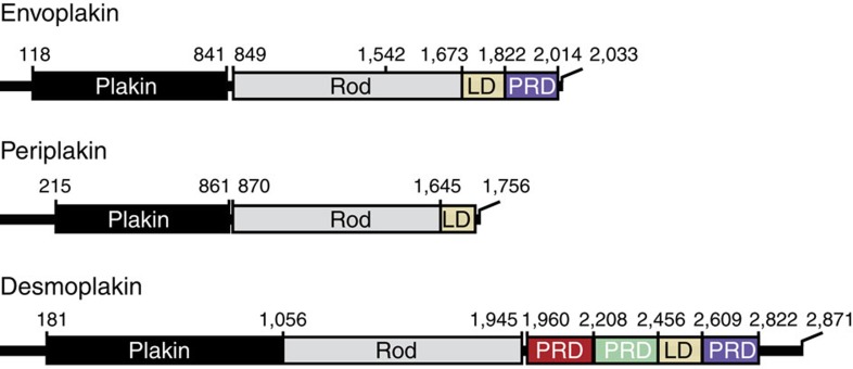
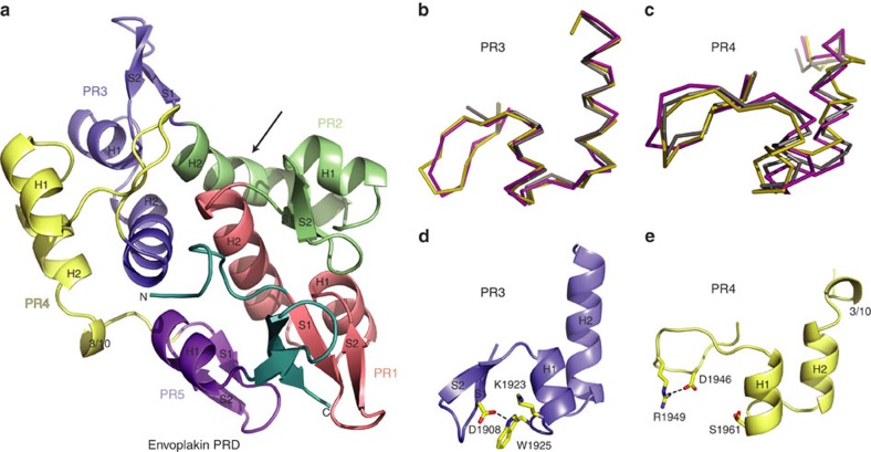
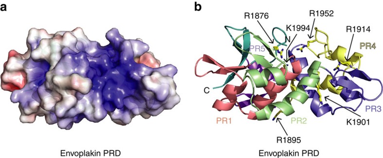
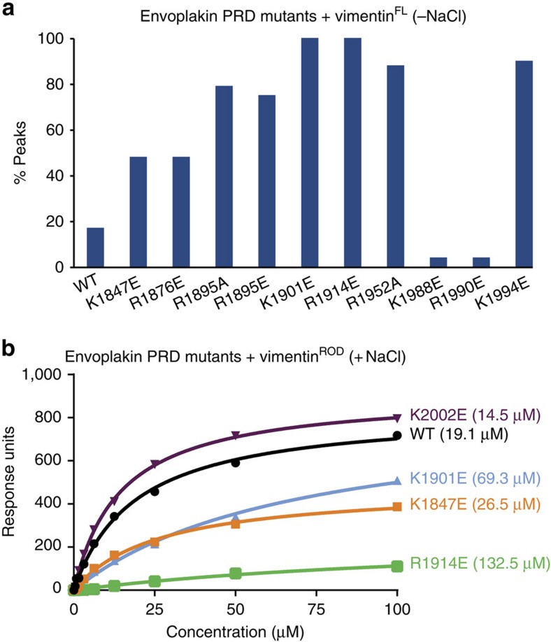
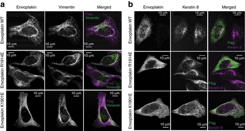
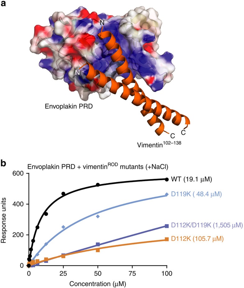
Plakin proteins form critical connections between cell junctions and the cytoskeleton; their disruption within epithelial and cardiac muscle cells cause skin-blistering diseases and cardiomyopathies. Envoplakin has a single plakin repeat domain (PRD) which recognizes intermediate filaments through an unresolved mechanism. Herein we report the crystal structure of envoplakin's complete PRD fold, revealing binding determinants within its electropositive binding groove. Four of its five internal repeats recognize negatively charged patches within vimentin via five basic determinants that are identified by nuclear magnetic resonance spectroscopy. Mutations of the Lys1901 or Arg1914 binding determinants delocalize heterodimeric envoplakin from intracellular vimentin and keratin filaments in cultured cells. Recognition of vimentin is abolished when its residues Asp112 or Asp119 are mutated. The latter slot intermediate filament rods into basic PRD domain grooves through electrosteric complementarity in a widely applicable mechanism. Together this reveals how plakin family members form dynamic linkages with cytoskeletal frameworks.


Figures

Figure 1
Figure 1. Domain architecture of plakin family members.
Envoplakin contains an N-terminal plakin domain, a central rod domain that forms a helical coiled coil, and a C-terminal tail region consisting of a linker domain (LD) and a single plakin repeat domain (PRD). Periplakin lacks a PRD module whereas desmoplakin possesses three. The domain boundaries are indicated.
Figure 2
Figure 2. Distinguishing features of the envoplakin PRD structure.
(a) Ribbon representation of the envoplakin PRD. Plakin repeats 1–5 are coloured red, green, blue, yellow and purple, respectively, with their PR motifs and secondary structural elements labelled. The N and C termini are labelled and shown in teal. The arrow indicates the kink of H2 in PR2. (b) Overlay of PR3 from envoplakin (magenta) and desmoplakin PRDs B (olive) and C (grey). (c) Overlay of PR4 from envoplakin and desmoplakin PRDs B and C. (d) Stabilization of the envoplakin PR3 by hydrogen bonding (black-dashed lines). (e) Stabilization of PR4 by hydrogen bonding.
Figure 3
Figure 3. The envoplakin PRD contains a conserved basic groove.
(a) Electrostatic surface potential of envoplakin PRD calculated with DelPhi. The potential scale ranges from −7 (red) to +7 (blue) in units of kT/e. (b) Ribbon representation of the envoplakin PRD highlighting the position of positively charged residues mutated in this study. The orientation is the same as in a, and the colours as in Fig. 2.
Figure 4
Figure 4. Mutating residues in envoplakin's basic groove abolishes vimentin binding.
(a) Histogram showing the percentage of 1H,15N amide peaks retaining more than 20% of their peak intensity on addition of 50 μM full-length vimentin to wild-type (WT) and mutant envoplakin PRD proteins (100 μM) in the absence of salt. (b) SPR analysis of wild-type and mutant envoplakin PRD binding to vimentinROD in the presence of 150 mM NaCl. The figures in parentheses are KD values. The data shown is representative of a number of experiments. KD values were estimated to be: WT=19.1±1.3 μM, K1847E=26.5±2.0 μM, K1901E=69.3±10.6 μM, R1914E=132.5±34.7 μM and K2002E=14.5±0.4 μM.
Figure 5
Figure 5. Intracellular distribution is compromised on mutating envoplakin's basic groove residues.
Constructs encoding residues 1542–2014 of human envoplakin with a C-terminal FLAG tag and residues 1588–1756 of human periplakin with a C-terminal HA tag (Supplementary Fig. 6a) were co-transfected into HeLa cells. The cells were stained with anti-FLAG (against envoplakin) and either (a) anti-vimentin or (b) anti-keratin 8 antibodies, showing the loss of cytoskeletal localization caused by the R1914E and K1901E mutations.
Figure 6
Figure 6. Modelling and binding studies of the envoplakin PRD–vimentin complex.
(a) The envoplakin PRD structure (coloured as in Fig. 3) was docked with vimentin Asn102-Leu138 (orange) (PDB 3G1E) using HADDOCK. (b) SPR analysis of wild-type envoplakin PRD binding to wild-type and mutant vimentinROD in the presence of 150 mM NaCl. The figures in parentheses are KD values in μM. The data shown is representative of a number of experiments. KD values were estimated to be: WT=19.1±1.3 μM, D112K=105.7±45.6 μM, D119K=48.4±5.5 μM and D112K/D119K=1505±139 μM.
Figure 7
Figure 7. Structural and functional relatedness of human PRDs.
(a) Phylogenetic tree of the PRD family including human envoplakin, desmoplakin, BPAG1e, BPAG1b, MACF1b, plectin and epiplakin, as generated by ClustalW. PRD modules are grouped into four subtypes, including a new PRD-D subtype. (b) Domain organization of the human plakin proteins with their PRD subtype and additional modules indicated. Note that BPAG1b and BPAG1e are encoded by the dystonin gene.

References

    1. Candi E., Schmidt R. & Melino G. The cornified envelope: a model of cell death in the skin. Nat. Rev. Mol. Cell Biol. 6, 328–340 (2005). - PubMed
    1. Sonnenberg A. & Liem R. K. Plakins in development and disease. Exp. Cell Res. 313, 2189–2203 (2007). - PubMed
    1. Huang Y., Li J. & Zhu X. Detection of anti-envoplakin and anti-periplakin autoantibodies by ELISA in patients with paraneoplastic pemphigus. Arch. Dermatol. Res. 301, 703–709 (2009). - PubMed
    1. Li J., Bu D. F., Huang Y. C. & Zhu X. J. Role of autoantibodies against the linker subdomains of envoplakin and periplakin in the pathogenesis of paraneoplastic pemphigus. Chin. Med. J. (Engl.) 122, 486–495 (2009). - PubMed
    1. Al-Jassar C., Bikker H., Overduin M. & Chidgey M. Mechanistic basis of desmosome-targeted diseases. J. Mol. Biol. 425, 4006–4022 (2013). - PMC - PubMed

Publication types