Review
. 2016 Jun;17(6):593-614.
doi: 10.1111/tra.12389. Epub 2016 Apr 7.

Fusion of Enveloped Viruses in Endosomes

Affiliations
Review

Fusion of Enveloped Viruses in Endosomes

Judith M White et al. Traffic. 2016 Jun.

Abstract

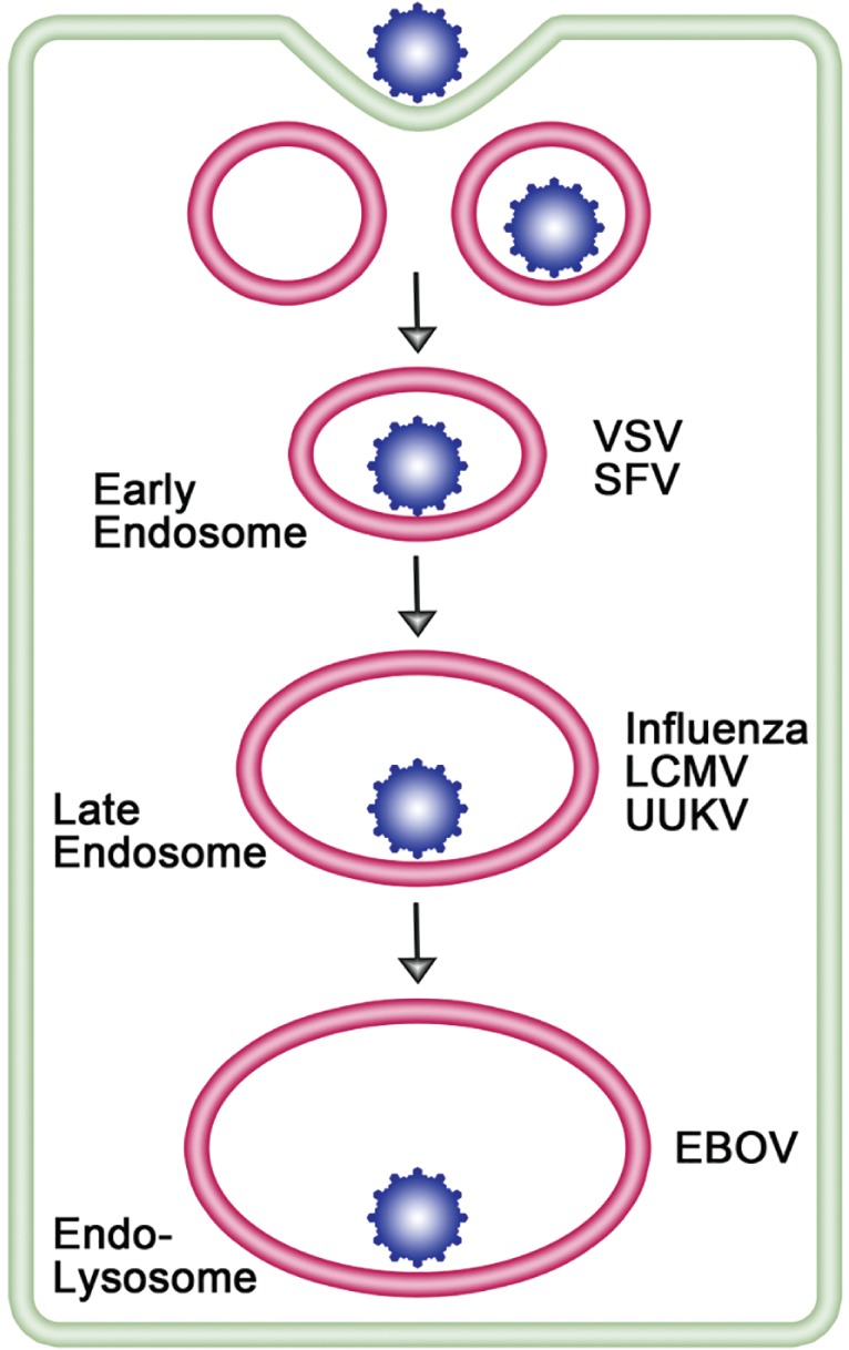
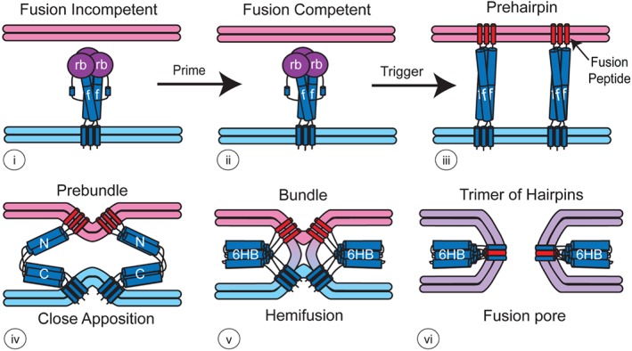
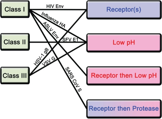
Ari Helenius launched the field of enveloped virus fusion in endosomes with a seminal paper in the Journal of Cell Biology in 1980. In the intervening years, a great deal has been learned about the structures and mechanisms of viral membrane fusion proteins as well as about the endosomes in which different enveloped viruses fuse and the endosomal cues that trigger fusion. We now recognize three classes of viral membrane fusion proteins based on structural criteria and four mechanisms of fusion triggering. After reviewing general features of viral membrane fusion proteins and viral fusion in endosomes, we delve into three characterized mechanisms for viral fusion triggering in endosomes: by low pH, by receptor binding plus low pH and by receptor binding plus the action of a protease. We end with a discussion of viruses that may employ novel endosomal fusion-triggering mechanisms. A key take-home message is that enveloped viruses that enter cells by fusing in endosomes traverse the endocytic pathway until they reach an endosome that has all of the environmental conditions (pH, proteases, ions, intracellular receptors and lipid composition) to (if needed) prime and (in all cases) trigger the fusion protein and to support membrane fusion.

Keywords: enveloped virus; fuse; low pH; membrane; prime; proteases; trigger; viral fusion protein; virus entry; virus receptors.


Figures

Figure 1
Figure 1
Enveloped virus entry through different endosomal compartments. Most enveloped viruses that enter the cell via endocytosis traverse the normal endocytic pathway (early endosome to late endosomes to endolysosome) and exit, by membrane fusion, where the conditions are sufficient to trigger the viral fusion protein; in some cases, the viral fusion protein is also proteolytically primed in the endocytic pathway as a prerequisite to fusion. [LCMV particles were found, however, to bypass early endosomes and traffic directly to, and fuse in, late endosomes 11, 12.] Examples of enveloped viruses that exit through early endosomes, late endosomes and endolysosomes are indicated. Viruses that enter through late endosomes or endolysosomes are termed ‘late penetrating viruses’ 13. See text and table legends for abbreviations.
Figure 2
Figure 2
Model for how viral fusion proteins function. The model shown is for a class I fusion protein, but related models apply to class II and III fusion proteins. The term for the state of the protein is given above each image. For most class I fusion proteins [see 67 for paramyxovirus F proteins], prior to triggering (i and ii), the receptor‐binding subunit (deep purple, rb) clamps the fusion subunit (dark blue, f). Upon triggering, the receptor‐binding subunit moves out of the way unclamping the fusion subunit so that it can form a prehairpin embedded in the target membrane via the fusion peptide (red). The prehairpin then folds back causing the N‐ and C‐α‐helical heptad repeats to form a six‐helix bundle (6HB) and progressively pulling the target (pink) and viral (light blue) membranes through stages of close apposition (iv), hemifusion (v) and fusion pore formation (vi). In some cases (e.g. for influenza HA), membrane coalescence is aided by further packing of sequences C‐terminal to the C‐heptad in the grooves of the central N‐heptad coiled coil 72. Importantly, for all characterized viral fusion proteins, the final (postfusion) conformation (vi) is a trimer‐of‐hairpins.
Figure 3
Figure 3
Different mechanisms by which class I, II and III fusion proteins are triggered. The three known classes of viral fusion proteins and the four confirmed mechanisms for fusion protein activation are shown on the left and right sides, respectively. Among fusion‐triggering mechanisms (right), blue denotes events that occur at neutral, and pink denotes ones that require low, pH. Some receptor + protease mechanisms do, whereas others do not, require low pH. Lines join ways in which specific viral fusion proteins, from different structural classes, are triggered. See text for details and abbreviations.

References

    1. Helenius A, Kartenbeck J, Simons K, Fries E. On the entry of Semliki forest virus into BHK‐21 cells. J Cell Biol 1980;84:404–420. - PMC - PubMed
    1. Matlin KS, Reggio H, Helenius A, Simons K. Infectious entry pathway of influenza virus in a canine kidney cell line. J Cell Biol 1981;91:601–613. - PMC - PubMed
    1. Marsh M, Helenius A. Adsorptive endocytosis of Semliki Forest virus. J Mol Biol 1980;142:439–454. - PubMed
    1. Marsh M, Bolzau E, Helenius A. Penetration of Semliki Forest virus from acidic prelysosomal vacuoles. Cell 1983;32:931–940. - PubMed
    1. Simons K, Garoff H, Helenius A. How an animal virus gets into and out of its host cell. Sci Am 1982;246:58–66. - PubMed

Publication types

Substances