. 2016 Apr;13(4):2999-3008.
doi: 10.3892/mmr.2016.4919. Epub 2016 Feb 22.

Collagen metabolic disorder induced by oxidative stress in human uterosacral ligament‑derived fibroblasts: A possible pathophysiological mechanism in pelvic organ prolapse

Affiliations

Collagen metabolic disorder induced by oxidative stress in human uterosacral ligament‑derived fibroblasts: A possible pathophysiological mechanism in pelvic organ prolapse

Cheng Liu et al. Mol Med Rep. 2016 Apr.

Abstract

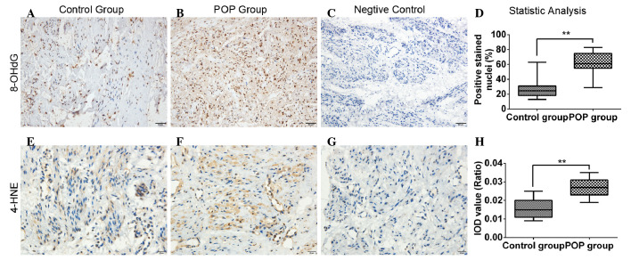
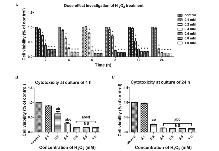
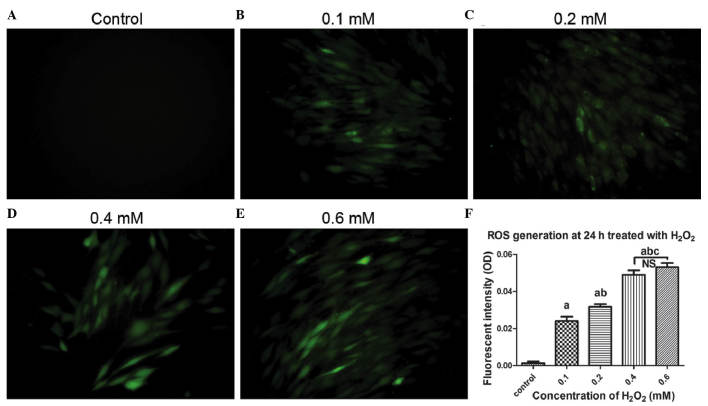
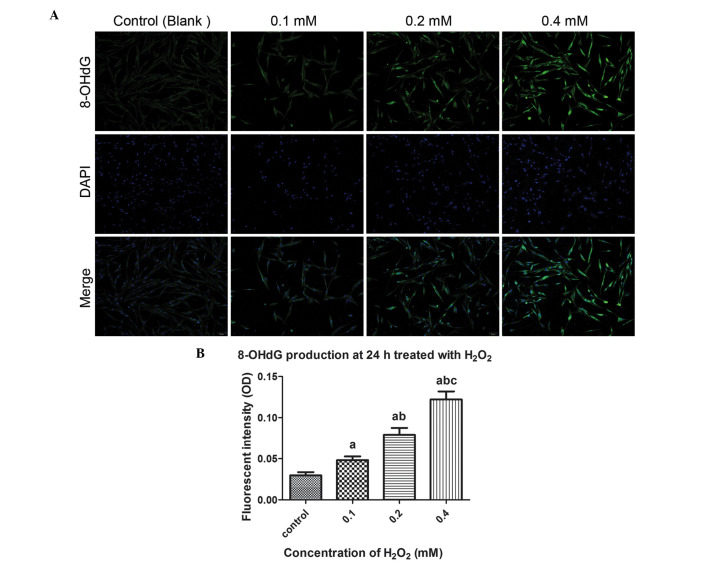
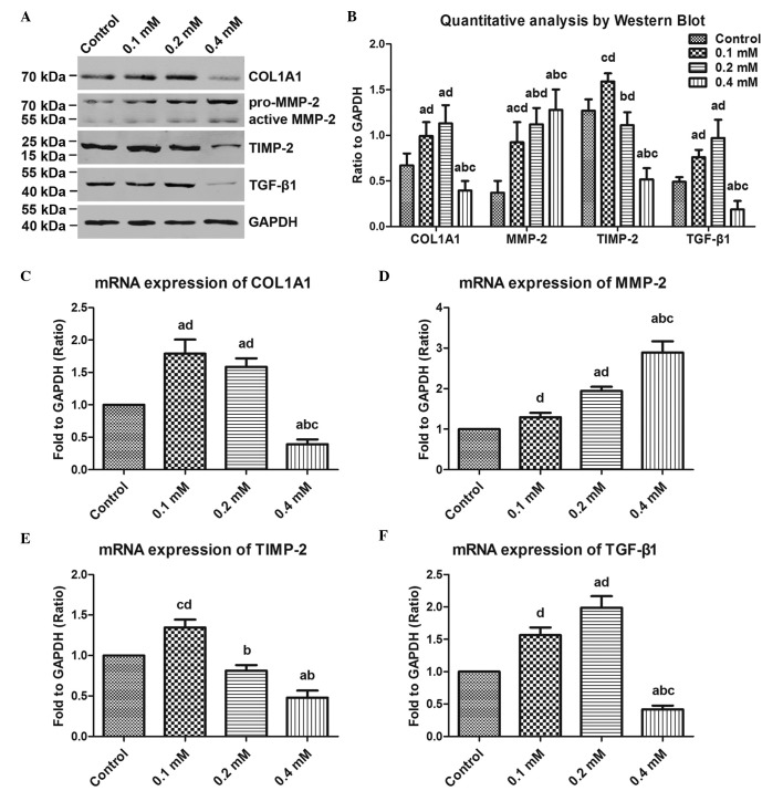
Pelvic organ prolapse (POP) is a global health problem, for which the pathophysiological mechanism remains to be fully elucidated. The loss of extracellular matrix protein has been considered to be the most important molecular basis facilitating the development of POP. Oxidative stress (OS) is a well‑recognized mechanism involved in fiber metabolic disorders. The present study aimed to clarify whether OS exists in the uterosacral ligament (USL) with POP, and to investigate the precise role of OS in collagen metabolism in human USL fibroblasts (hUSLFs). In the present study, 8‑hydroxyguanosine (8‑OHdG) and 4 hydroxynonenal (4‑HNE), as oxidative biomarkers, were examined by immunohistochemistry to evaluate oxidative injury in USL sections in POP (n=20) and non‑POP (n=20) groups. The primary cultured hUSLFs were treated with exogenous H2O2 to establish an original OS cell model, in which the expression levels of collagen, type 1, α1 (COL1A1), matrix metalloproteinase (MMP)‑2, tissue inhibitor of metalloproteinase (TIMP)‑2 and transforming growth factor (TGF)‑β1 were evaluated by western blot and reverse transcription‑quantitative polymerase chain reaction analyses. The results showed that the expression levels of 8‑OHdG and 4‑HNE in the POP group were significantly higher, compared with those in the control group. Collagen metabolism was regulated by H2O2 exposure in a concentration‑dependent manner, in which lower concentrations of H2O2 (0.1‑0.2 mM) stimulated the anabolism of COL1A1, whereas a higher concentration (0.4 mM) promoted catabolism. The expression levels of MMP‑2, TIMP‑2 and TGF‑β1 exhibited corresponding changes with the OS levels. These results suggested that OS may be involved in the pathophysiology of POP by contributing to collagen metabolic disorder in a severity‑dependent manner in hUSLFs, possibly through the regulation of MMPs, TIMPs and TGF‑β1 indirectly.


Figures

Figure 1
Figure 1
Immunohistochemical analysis for assessment of rgw immunoreactivity of 8-OHdG and 4-HNE in uterosacral ligaments. (A–C) Immunohistochemical staining for 8-OHdG, (magnification, ×200; scale bar=50 µm). (D) Statistical analysis, based on the percentage of 8-OHdG-positive stained cells in the total cell count. (E–G) Immunohistochemical staining for 4-HNE (magnification, ×400; scale bar=20 µm). (H) Statistical analysis, based on the IOD value. Data are presented as the mean ± standard deviation (**P<0.01). 8-OHdG, 8-hydroxyguanosine; 4-HNE, 4-hydroxynonenal; IOD, integrated optical density; POP, pelvic organ prolapse.
Figure 2
Figure 2
Effects of exogenous H2O2 on the cell viability of human uterosacral ligament fibroblasts. Cell viability was examined using a Cell Counting Kit-8 assay and data are presented as apercentage of the untreated control group. (A) Cells were treated with the indicated concentrations of H2O2 for 2, 4, 6, 8, 12 and 24 h for determination of concentration-dependence. Cell viability decreased in a concentration-dependent and time-dependent manner under H2O2 exposure. *P<0.05, vs. control group (two-way ANOVA. The cells were treated with the indicated concentrations of H2O2 for (B) 4 h and (C) 24 h for determination of concentration-dependence. One-way ANOVA was performed, and data are presented as the median ± standard error of the mean (n=3). a, P<0.05, vs. unstreated control; b, P<0.05, vs. 0.1 mM; c, P<0.05, vs. 0.2 mM; d, P<0.05, vs. 0.4 mM; NS, no significance; ANOVA, analysis of variance; H2O2, hydrogen peroxide.
Figure 3
Figure 3
Cell apoptosis is induced by exogenous H2O2. Following treatment with different concentrations of H2O2 for 24 h, the human uterosacral ligament fibroblasts were stained with Annexin V/propidium iodide and then assayed using flow cytometry. Cell apoptosis following exposure to (A) 0, (B) 0.1, (C) 0.2, (D) 0.4 and (E) 0.6 mM H2O2. (F) Statistical analysis of the apoptotic rates. Data are presented as the median ± standard error of the mean. One-way analysis of variance was performed, followed by an unpaired t-test. Data are presented as the median ± standard error of the mean (n=3). a, P<0.05, vs. untreated control; b, P<0.05, vs. 0.1 mM; c, P<0.05, vs. 0.2 mM; NS, no significance; H2O2, hydrogen peroxid; PI, propidium iodide; FITC, fluorescein isothocyanate.
Figure 4
Figure 4
Microscopic images of ROS generation induced by H2O2 treatment using DCF-DA staining. The cells were pre-treated with exogenous H2O2 at concentrations of (A) 0, (B) 0.1, (C) 0.2, (D) 0.4 and (E) 0.6 mM, and then incubated with DCF-DA. The cells were observed under a fluorescent microscope (magnification, ×200). (F) Quantitative analysis based on fluorescence intensity, obtained using Image-pro Plus 6.0 software. Data are presented as the median ± standard error of the mean. One-way analysis of variance was performed, followed by an unpaired t-test. Data are presented as the median ± standard error of the mean (n=3). a, P<0.05, vs. untreated control; b, P<0.05, vs. 0.1 mM; c, P<0.05, vs. 0.2 mM; NS, no significance; ROS, reactive oxygen species; OD, optical density; H2O2, hydrogen peroxide; DCF-DA, 2′,7′-dichlorofluorescein diacetate.
Figure 5
Figure 5
Microscopic images of 8-OHdG production induced by H2O2 treatment using an immunofluorescent assay. (A) Cells were pre-treated with exogenous H2O2 (0, 0.1, 0.2, and 0.4 mM) for 24 h, followed by incubation with 8-OHdG antibodies and staining with DAPI. The cells were observed under a fluorescent microscope (magnification, ×200). (B) Quantitative analysis based on fluorescence intensity, obtained using image-pro plus 6.0 software. One-way analysis of variance was performed, followed by an unpaired t-test. Data are presented as the median ± standard error of the mean (n=3). a, P<0.05, vs. untreated control; b, P<0.05, vs. 0.1 mM; c, P<0.05, vs. 0.2 mM; NS, no significance; 8-OHdg, 8-hydroxyguanosine; OD, optical density; H2O2, hydrogen peroxide.
Figure 6
Figure 6
Effects of exogenous H2O2 on COL1A1 metabolism in human uterosacral ligament fibroblasts. The cells were pre-treated with the indicated concentrations of H2O2 (0, 0.1, 0.2 and 0.4 mM) for 24 h, and then assayed by Western blot and RT-qPCR analyses. (A) COL1A1, MMP-2, TIMP-2 and TGF-β1 were examined using Western blot analysis at the protein level. (B) Quantitative analysis based on the bands of the Western blot. The mRNA expression levels of (C) COL1A1, (D) MMP-2, (E) TIMP-2 and (F) TGF-β1 were determined by RT-qPCR. One-way analysis of variance was performed and data are presented as the median ± standard error of the mean (n=3). a, P<0.05, vs. untreated control; b, P<0.05, vs. 0.1 mM; c, P<0.05, vs. 0.2 mM; d, P<0/05, vs. 0.4 mM; NS, no significance; COL1A1, collagen, type 1, α1; MMP-2, matrix metalloproteinase-2, TIMP-2, tissue inhibitor of metalloproteinase-2; TGF-β1, transforming growth factor-β1; H2O2, hydrogen peroxide.

References

    1. Barber MD, Maher C. Epidemiology and outcome assessment of pelvic organ prolapse. Int Urogynecol J. 2013;24:1783–1790. doi: 10.1007/s00192-013-2169-9. - DOI - PubMed
    1. Olsen AL, Smith VJ, Bergstrom JO, Colling JC, Clark AL. Epidemiology of surgically managed pelvic organ prolapse and urinary incontinence. Obstet Gynecol. 1997;89:501–506. doi: 10.1016/S0029-7844(97)00058-6. - DOI - PubMed
    1. Subak LL, Waetjen LE, van den Eeden S, Thom DH, Vittinghoff E, Brown JS. Cost of pelvic organ prolapse surgery in the United States. Obstet Gynecol. 2001;98:646–651. - PubMed
    1. Merrill RM. Hysterectomy surveillance in the United States, 1997 through 2005. Med Sci Monit. 2008;14:CR24–CR31. - PubMed
    1. Weber AM, Buchsbaum GM, Chen B, Clark AL, Damaser MS, Daneshgari F, Davis G, DeLancey J, Kenton K, Weidner AC, Word RA. Basic science and translational research in female pelvic floor disorders: Proceedings of an NIH-sponsored meeting. Neurourol Urodyn. 2004;23:288–301. doi: 10.1002/nau.20048. - DOI - PubMed

Publication types

MeSH terms