. 2016 Jun;41(5):415-25.
doi: 10.1093/chemse/bjw016. Epub 2016 Mar 2.

The Pig Olfactory Brain: A Primer

Affiliations

The Pig Olfactory Brain: A Primer

Peter C Brunjes et al. Chem Senses. 2016 Jun.

Abstract

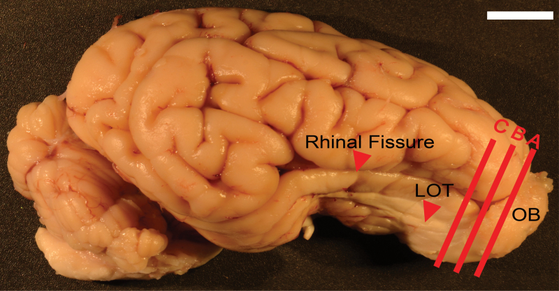
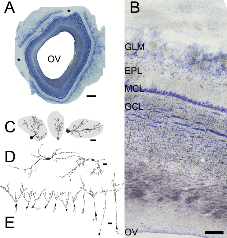
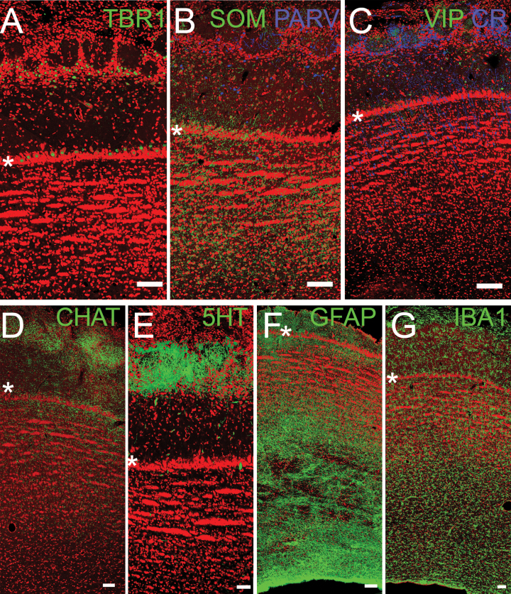
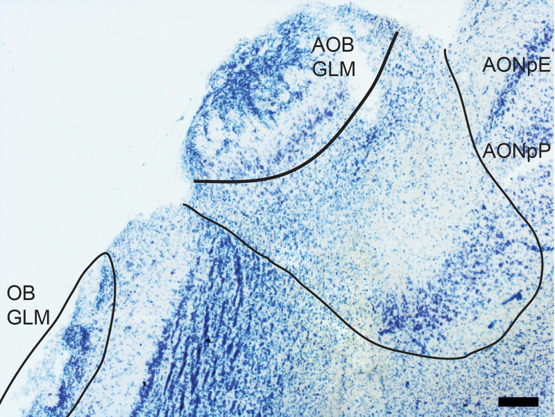
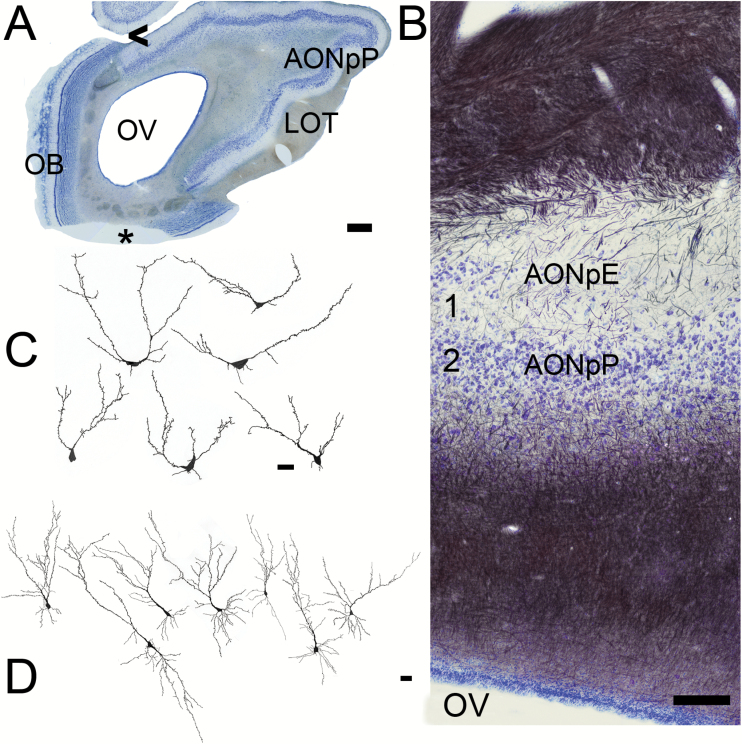
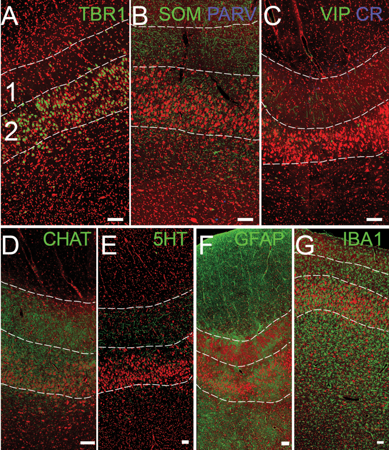
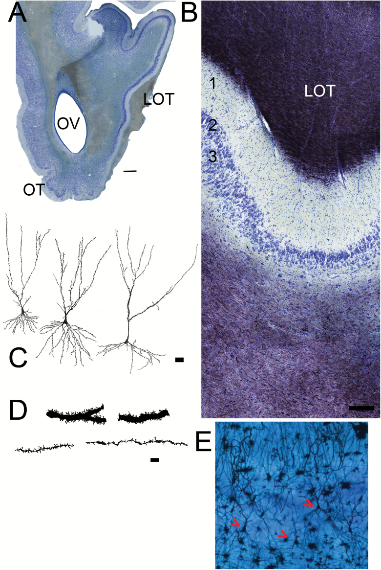
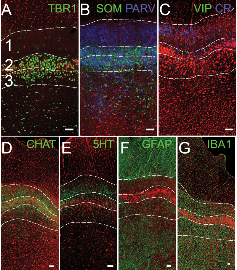
Despite the fact that pigs are reputed to have excellent olfactory abilities, few studies have examined regions of the pig brain involved in the sense of smell. The present study provides an overview of the olfactory bulb, anterior olfactory nucleus, and piriform cortex of adult pigs using several approaches. Nissl, myelin, and Golgi stains were used to produce a general overview of the organization of the regions and confocal microscopy was employed to examine 1) projection neurons, 2) GABAergic local circuit neurons that express somatostatin, parvalbumin, vasoactive intestinal polypeptide, or calretinin, 3) neuromodulatory fibers (cholinergic and serotonergic), and 4) glia (astrocytes and microglia). The findings revealed that pig olfactory structures are quite large, highly organized and follow the general patterns observed in mammals.

Keywords: anterior olfactory nucleus; olfactory bulb; olfactory cortex; olfactory peduncle; piriform cortex.


Figures

Figure 1.
Figure 1.
Pig brain from the lateral side, anterior to right and dorsal to top. A, B, C = approximate planes of section for Nissl sections in Figures 2A, 5A and 7A, respectively. Red arrow delineates the rhinal fissure, which separates the olfactory cortex (ventral) from the cerebral cortex (dorsal). Scale bar = 1cm.
Figure 2.
Figure 2.
Sections from the OB. (A) Low magnification picture (scale bar = 1mm) showing the large olfactory ventricle (OV) and the easily recognizable laminar structure of the bulb. Asterisks = missing portions. (B) Nissl (blue)/myelin (black) staining. Superficial at top, the OV at bottom. The characteristic layers of the bulb include the glomerular layer (GLM), with its spherical regions of neuropil, the broad, relatively cell free EPL, compact MCL, and striated GCL. Scale bar = 200 µm. (C, D) Camera lucida drawings of Golgi–Cox stained cells, scale bars = 25 µm. (C) Periglomerular cells, whose dendrites extend into glomeruli (grey) and their somata are found in region between the glomeruli. (D) Large, horizontal cells from the EPL. (E) Granule cells, whose dendritic tufts are located in the EPL and somata in the superficial (left) to deep (right) GCL. The cell body of the left-most granule cell was located deep within the MCL.
Figure 3.
Figure 3.
Confocal images of OB. Asterisks delineate the MCL for orientation (Figure 2B). (A) TBR1 staining (green) labels projection neurons: mitral and tufted cells. (B, C) Interneurons stained for SOM, PARV, VIP, and CR. (D, E) Neuromodulatory inputs from higher brain regions, including cholinergic (CHAT) and serotonergic (5-HT) fibers. (F, G) Glial cells including astrocytes (GFAP) and microglia (IBA-1). Red = Nissl stain. Scale bars = 100µm
Figure 4.
Figure 4.
The accessory OB. This structure receives sensory input from the vomeronasal organ via the vomeronasal nerve, and is located on the dorsomedial surface of the caudal OB. The structure can be found just medial to the region in which the olfactory nerve and glomerular layer thin and then disappear. Medial to the AOB, the 2 regions that form the AON (AONpE) and AONpP emerge. Scale bar = 30 µm.
Figure 5.
Figure 5.
Sections from the AON. (A) Low magnification picture (scale bar = 1mm). The OB is still present on the medial (left) side of the olfactory peduncle. The LOT is visible on the right (lateral) side. AONpP fills the remainder of the region on the lateral side. OV = olfactory ventricle. Asterisk = missing portion. (B) Nissl (blue)/myelin (black) staining. Superficial at top, the OV at bottom. The top of the section contains myelinated fibers and collaterals from the LOT. They thin toward the ventral side, indicating the beginning the Layer 1 (1A) of the AONpP. Under the cell poor Layer 1 is the cellular region, Layer 2. Beneath Layer 2 is a second large region of fibers that includes the axons of AON projection neurons and afferents from higher brain centers. Within Layer 1 AONpE can also be seen as a thin band of cells at the left side of the figure. Arrowhead = area where AOB is found in more rostral sections. Scale bar = 200 µm. (C, D) Camera lucida drawings of Golgi-stained cells. Scale bars = 50 µm. (C) AONpE cells. These characteristic cells have only a few apically directed dendrites arising from the cell body. (D) AONpP pyramidal cells. There neurons have a single apically directed dendrite (at top) and several basilar ones.
Figure 6
Figure 6
Confocal images of AONpP. The superficial side (pial) is toward the top, the olfactory ventricle to the bottom. The numbers 1 and 2 in (A) and dotted lines delineate the approximate boundaries of layers 1 and 2 for orientation (Figure 5B). (A) TBR1 staining (green) labels projection neurons: pyramidal cells. (B, C) Interneurons stained for SOM, PARV, VIP, and CR. (D, E) Neuromodulatory inputs from higher brain regions, including cholinergic (CHAT) and serotonergic (5-HT) fibers. (F, G) Glial cells, including astrocytes (GFAP) and microglia (IBA-1). Red = Nissl stain. Scale bar = 100 µm.
Figure 7.
Figure 7.
Sections from the anterior PC. (A) Low magnification picture (scale bar = 1mm). The olfactory tubercle has emerged on the ventral side of the olfactory peduncle. The LOT is visible on the right (lateral) side. The APC occupies the entire lateral side of the peduncle. (B) Nissl (blue)/myelin (black) staining. Superficial at top. As in Figure 5, the top of the section contains myelinated fibers and collaterals from the LOT that thin toward the deeper regions indicating the beginning the Layer 1 (1A) of the APC. Under the cell poor Layer 1 are cellular layers 2 and 3. Scale bar = 200 µm. (C) Camera lucida drawings of pyramidal neurons from the APC. Scale bar = 50 µm. (D) Higher magnification drawings of sections of apical dendrites from APC pyramidal neurons. Some cells have large caliber dendrites with many spines (top), others have much finer processes with fewer spines (bottom). Scale bar = 10 µm. (E) Photomicrograph of Golgi/Nissl section showing three pyramidal neurons (red arrows).
Figure 8.
Figure 8.
Confocal images of the anterior PC. The superficial side (pial) is toward the top. The numbers 1, 2, and 3 in panel (A) and dotted lines delineate the approximate borders of layers 1–3 for orientation (Figure 7B). (A) TBR1 staining (green) labels projection neurons: pyramidal cells. (B, C) Interneurons stained for SOM, PARV, VIP, and CR. (D, E) Neuromodulatory inputs from higher brain regions, including cholinergic (CHAT) and serotonergic (5-HT) fibers. (F, G) Glial cells including astrocytes (GFAP) and microglia (IBA-1). Red = Nissl stain. Scale bars = 100 µm.

References

    1. Amaral AJ, Ferretti L, Megens HJ, Crooijmans RP, Nie H, Ramos-Onsins SE, Perez-Enciso M, Schook LB, Groenen MA. 2011. Genome-wide footprints of pig domestication and selection revealed through massive parallel sequencing of pooled DNA. PLoS One. 6(4):e14782. - PMC - PubMed
    1. Baldwin BA. 1980. Operant studies and preference tests on the role of olfaction and taste in the ingestive behaviour of pigs and ruminants. Fortschr Tierphysiol Tierernahr. (11):36–42. - PubMed
    1. Bekkers JM, Suzuki N. 2013. Neurons and circuits for odor processing in the piriform cortex. Trends Neurosci. 36(7):429–438. - PubMed
    1. Booth W, Signoret J. 1992. Olfaction and reproduction in ungulates. Oxf Rev Reprod Biol. 14:263–301. - PubMed
    1. Booth WD, Baldwin BA. 1980. Lack of effect on sexual behaviour or the development of testicular function after removal of olfactory bulbs in prepubertal boars. J Reprod Fertil. 58(1):173–182. - PubMed

Publication types

LinkOut - more resources