

Longitudinal diffusion MRI for treatment response assessment: Preliminary experience using an MRI-guided tri-cobalt 60 radiotherapy system
- PMID: 26936721
- PMCID: PMC6961701
- DOI: 10.1118/1.4942381
Longitudinal diffusion MRI for treatment response assessment: Preliminary experience using an MRI-guided tri-cobalt 60 radiotherapy system
Abstract
Purpose: To demonstrate the preliminary feasibility of a longitudinal diffusion magnetic resonance imaging (MRI) strategy for assessing patient response to radiotherapy at 0.35 T using an MRI-guided radiotherapy system (ViewRay).
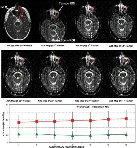
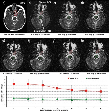
Methods: Six patients (three head and neck cancer, three sarcoma) who underwent fractionated radiotherapy were enrolled in this study. A 2D multislice spin echo single-shot echo planar imaging diffusion pulse sequence was implemented on the ViewRay system and tested in phantom studies. The same pulse sequence was used to acquire longitudinal diffusion data (every 2-5 fractions) on the six patients throughout the entire course of radiotherapy. The reproducibility of the apparent diffusion coefficient (ADC) measurements was assessed using reference regions and the temporal variations of the tumor ADC values were evaluated.
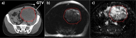
Results: In diffusion phantom studies, the ADC values measured on the ViewRay system matched well with reference ADC values with <5% error for a range of ground truth diffusion coefficients of 0.4-1.1 × 10(-3) mm(2)/s. The remote reference regions (i.e., brainstem in head and neck patients) had consistent ADC values throughout the therapy for all three head and neck patients, indicating acceptable reproducibility of the diffusion imaging sequence. The tumor ADC values changed throughout therapy, with the change differing between patients, ranging from a 40% drop in ADC within the first week of therapy to gradually increasing throughout therapy. For larger tumors, intratumoral heterogeneity was observed. For one sarcoma patient, postradiotherapy biopsy showed less than 10% necrosis score, which correlated with the observed 40% decrease in ADC from the fifth fraction to the eighth treatment fraction.
Conclusions: This pilot study demonstrated that longitudinal diffusion MRI is feasible using the 0.35 T ViewRay MRI. Larger patient cohort studies are warranted to correlate the longitudinal diffusion measurements to patient outcomes. Such an approach may enable response-guided adaptive radiotherapy.
Figures
References
-
- Malayeri A. A., El Khouli R. H., Zaheer A., Jacobs M. A., Corona‐Villalobos C. P., Kamel I. R., and Macura K. J., “Principles and applications of diffusion‐weighted imaging in cancer detection, staging, and treatment follow‐up,” RadioGraphics 31, 1773–1791 (2011). 10.1148/rg.316115515 - DOI - PMC - PubMed
-
- Il Yu J., Park H. C., Lim D. H., Choi Y., Jung S. H., Paik S. W., Kim S. H., Jeong W. K., and Kim Y. K., “The role of diffusion‐weighted magnetic resonance imaging in the treatment response evaluation of hepatocellular carcinoma patients treated with radiation therapy,” Int. J. Radiat. Oncol., Biol., Phys. 89, 814–821 (2014). 10.1016/j.ijrobp.2014.03.020 - DOI - PubMed
MeSH terms
Substances
Grants and funding
LinkOut - more resources
Full Text Sources
Other Literature Sources
