Review
. 2016 Mar;53(2):349-65.
doi: 10.1177/0300985815623621.

Endocrinopathy and Aging in Ferrets

Affiliations
Review

Endocrinopathy and Aging in Ferrets

V Bakthavatchalu et al. Vet Pathol. 2016 Mar.

Abstract

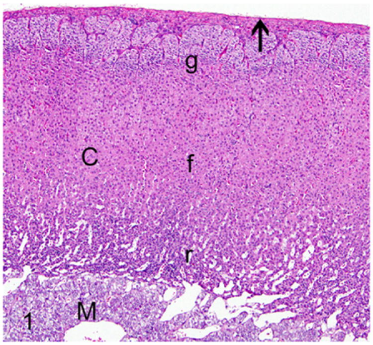
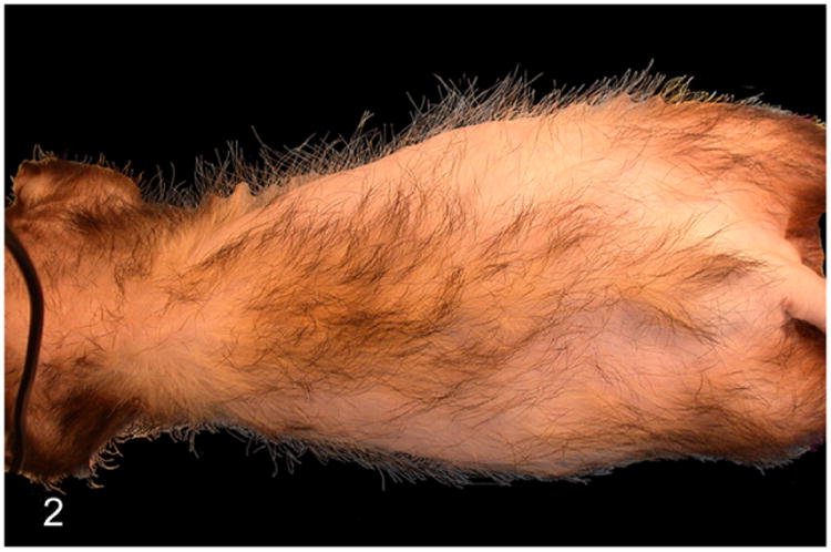
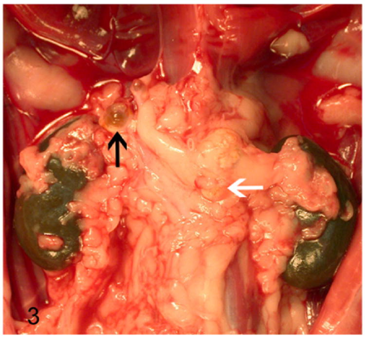
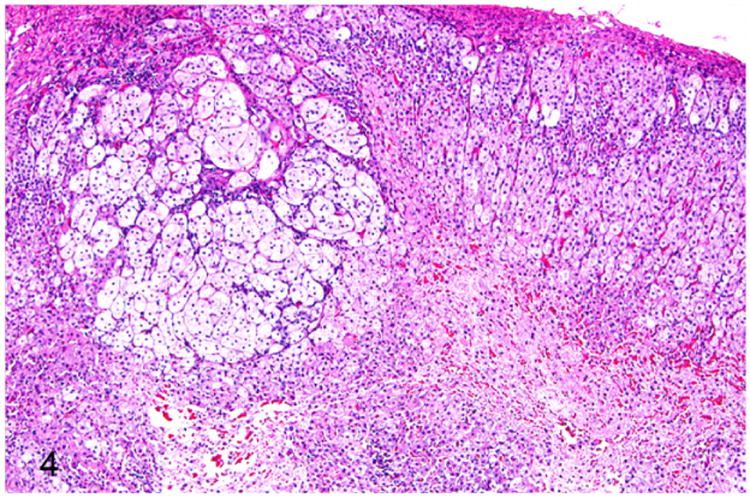
Ferrets have become more popular as household pets and as animal models in biomedical research in the past 2 decades. The average life span of ferrets is about 5-11 years with onset of geriatric diseases between 3-4 years including endocrinopathies, neoplasia, gastrointestinal diseases, cardiomyopathy, splenomegaly, renal diseases, dental diseases, and cataract. Endocrinopathies are the most common noninfectious disease affecting middle-aged and older ferrets. Spontaneous neoplasms affecting the endocrine system of ferrets appear to be increasing in prevalence with a preponderance toward proliferative lesions in the adrenal cortex and pancreatic islet cells. Diet, gonadectomy, and genetics may predispose ferrets to an increased incidence of these endocrinopathies. These functional proliferative lesions cause hypersecretion of hormones that alter the physiology and metabolism of the affected ferrets resulting in a wide range of clinical manifestations. However, there is an apparent dearth of information available in the literature about the causal relationship between aging and neoplasia in ferrets. This review provides a comprehensive overview of the anatomy and physiology of endocrine organs, disease incidence, age at diagnosis, clinical signs, pathology, and molecular markers available for diagnosis of various endocrine disorders in ferrets.

Keywords: adrenal-associated endocrinopathy; adrenocortical neoplasm; aging; and thyroid; cysts; diabetes mellitus; endocrine tumors; endocrinopathies; estrogen-induced anemia; ferret; gonadectomy; insulinoma; islet cell tumor; multiple endocrine neoplasia; neuroblastoma; ovary; pancreatic polypeptidoma; parathyroid; pheochromocytoma; pituitary; review; teratoma; testis.


Conflict of interest statement

Declaration of Conflicting Interests: The author(s) declared no potential conflicts of interest with respect to the research, authorship, and/or publication of this article.

Figures

Figure 1
Figure 1
Proliferative lesions of the adrenal gland, ferret. Normal adrenal gland. The adrenal gland has a dense fibrous capsule (arrow). The adrenal cortex (C) is divided into three zones: zona glomerulosa (g), zona fasciculata (f), and zona reticularis (r). HE, hematoxylin and eosin; M, medulla. Hematoxylin and eosin (HE).
Figure 2
Figure 2
Proliferative lesions of the adrenal gland, ferret. Adrenal-associated endocrinopathy. Bilaterally symmetrical dorsolateral truncal alopecia. The causative adrenocortical tumor is shown in Fig. 3.
Figure 3
Figure 3
Proliferative lesions of the adrenal gland, ferret. Adrenocortical tumor. Cystic right adrenal gland (black arrow) and an enlarged neoplastic left adrenal gland (white arrow) embedded within adipose tissue.
Figure 4
Figure 4
Proliferative lesions of the adrenal gland, ferret. Focal adrenocortical hyperplasia. The mass is formed by large clear cells with vacuolated cytoplasm. HE.
Figure 5
Figure 5
Proliferative lesions of the adrenal gland, ferret. Cysts of probable biliary origin, adrenal gland (from Fig. 3). The right adrenal gland and adjacent adipose tissue are disrupted by multiple variably sized cysts lined by attenuated low cuboidal epithelium and containing homogenous eosinophilic proteinaceous material. HE.
Figure 6
Figure 6
Proliferative lesions of the adrenal gland, ferret. Adrenocortical adenoma (from Fig. 3). Neoplastic adrenocortical cells compress adjacent normal adrenocortical tissue (top right).
Figure 7
Figure 7
Proliferative lesions of the adrenal gland, ferret. Adrenocortical carcinoma. The neoplasm is composed of clear cells with pale vacuolated cytoplasm and polygonal cells supported by fibrous connective tissue stroma. Hematoxylin and eosin (HE).
Figure 8
Figure 8
Proliferative lesions of the adrenal gland, ferret. Adrenocortical carcinoma with myxoid differentiation. Neoplastic cells often form cystic spaces that contain amphophilic to pale basophilic material (mucin). HE.
Figure 9
Figure 9
Proliferative lesions of the adrenal gland, ferret. Pheochromocytoma and cortical adenoma, adrenal gland. A well-demarcated expansile pheochromocytoma that compresses an adjacent adrenocortical adenoma (on the left). HE.
Figure 10
Figure 10
Proliferative lesions of the adrenal gland, ferret. Higher magnification of Fig. 9. Polyhedral neoplastic cells are arranged in nest and packets supported by fine fibrovascular stroma. HE.
Figure 11
Figure 11
Pancreatic islet cell neoplasm (insulinoma), ferret. Expansion and fusion of multiple proliferating neoplastic islets. HE.
Figure 12
Figure 12
Higher magnification image of Fig. 11. Cords of well-differentiated neoplastic islet cells palisade around blood vessels. HE.
Figure 13
Figure 13
Sex-cord stromal tumor, ovary, ferret. Ovarian architecture is effaced by a lobulated highly cellular spindle cell neoplasm supported by dense fibrous connective tissue. HE.
Figure 14
Figure 14
Higher magnification of Fig. 13. The biphasic neoplasm is composed of interlacing streams and bundles of spindle cells and polygonal cells arranged in cords. HE.
Figure 15
Figure 15
Sertoli cell tumor, testis, ferret. Tubular structures are composed of multiple layers of neoplastic cells. The neoplastic cells are arranged perpendicular to the basement membrane (inset). HE.
Figure 16
Figure 16
Higher magnification of Fig. 15. Neoplastic cells palisade around blood vessels. The elongated polygonal neoplastic cells have abundant eosinophilic granular cytoplasm with intracytoplasmic vacuoles. HE.
Figure 17
Figure 17
Interstitial cell tumor, testicle, ferret. An expansile poorly demarcated neoplasm compresses the adjacent seminiferous tubules (on the left). HE.
Figure 18
Figure 18
Higher magnification of Fig. 17. Sheets of well-differentiated large polygonal neoplastic cells contain abundant finely granular to vacuolated cytoplasm and large round nuclei. HE.
Figure 19
Figure 19
Lymphoplasmacytic thyroiditis, ferret. Severe disruption of thyroid architecture by perifollicular aggregates of lymphocytes and plasma cells. Follicles within the affected area are collapsed and contain minimal amounts of colloid admixed with cellular debris. HE.
Figure 20
Figure 20
Thyroid gland adenocarcinoma, ferret. Adapted from Wills et al. The neoplasm forms irregular follicles infiltrating adjacent muscle. Inset: The neoplastic follicular structures contain minimal amounts of colloid. HE.
Figure 21
Figure 21
Thyroid gland adenocarcinoma, ferret. Adapted from Wills et al. Neoplastic cells lining follicles demonstrate cytoplasmic thyroglobulin expression. Immunohistochemistry for thyroglobulin.
Figure 22
Figure 22
C-cell carcinoma, thyroid gland, ferret. An irregularly protruding mass on the ventrolateral aspect of the neck.
Figure 23
Figure 23
C-cell carcinoma, thyroid gland, ferret. The neoplasm is composed of polygonal cells arranged in nests, packets, and anastomosing cords. Neoplastic cells are separated by dense fibrovascular connective tissue and often form necrotic foci. HE.
Figure 24
Figure 24
Neoplastic cells have indistinct cell borders with scant vacuolated cytoplasm and oval to round nuclei. Neoplastic cells palisade around blood vessels. HE.

References

    1. Allanson M. The reproductive processes of certain mammals. III—the reproductive cycle of the male ferret. Proc Biol Sci. 1932;110(767):295–312.
    1. Andrews GA, Myers NC, Chard-Bergstrom C. Immunohistochemistry of pancreatic islet cell tumors in the ferret (Mustela putorius furo) Vet Pathol. 1997;34(5):387–393. - PubMed
    1. Antinoff N, Hahn K. Ferret oncology: diseases, diagnostics, and therapeutics. Vet Clin North Am Exot Anim Pract. 2004;7(3):579–625. - PubMed
    1. Antinoff N, Williams BH. Neoplasia. In: Quesenberry KE, Carpenter JW, editors. Ferrets, Rabbits, and Rodents. 3rd. Saint Louis, MO: W.B. Saunders; 2012. pp. 103–121.
    1. Ball RS. Issues to consider for preparing ferrets as research subjects in the laboratory. ILAR J. 2006;47(4):348–357. - PMC - PubMed

Publication types

LinkOut - more resources