Review
. 2015:80:93-101.
doi: 10.1101/sqb.2015.80.027193. Epub 2016 Mar 2.

A System to Study Aneuploidy In Vivo

Affiliations
Review

A System to Study Aneuploidy In Vivo

Sarah J Pfau et al. Cold Spring Harb Symp Quant Biol. 2015.

Abstract

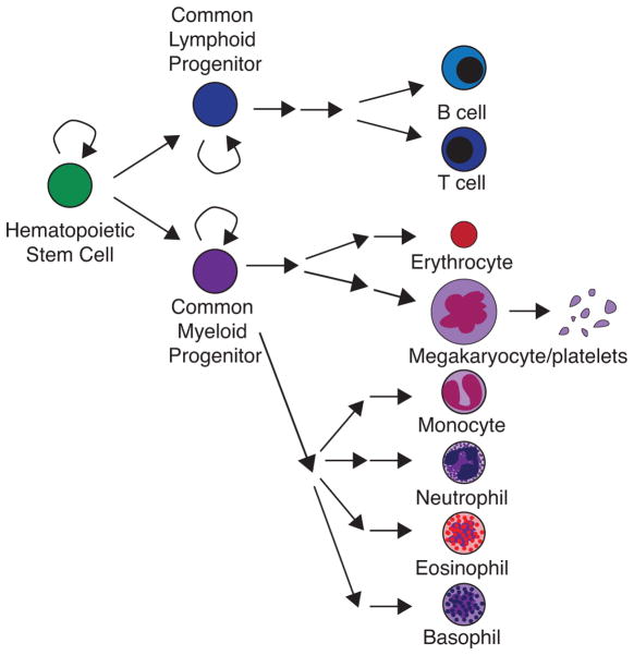
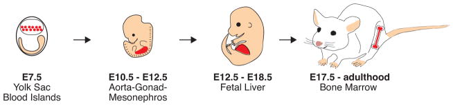
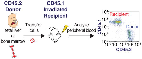
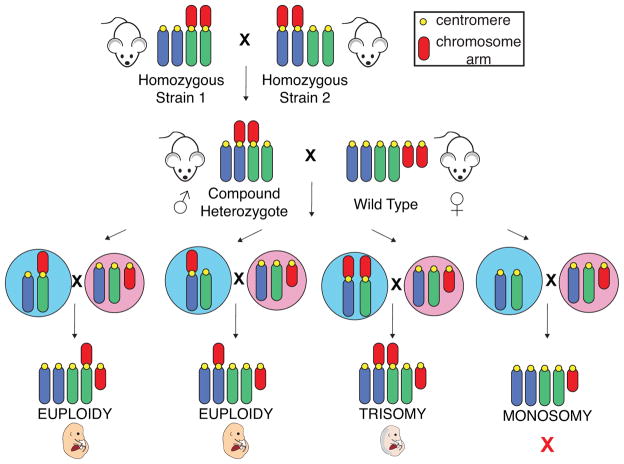
Aneuploidy, an imbalanced chromosome number, is associated with both cancer and developmental disorders such as Down syndrome (DS). To determine how aneuploidy affects cellular and organismal physiology, we have developed a system to evaluate aneuploid cell fitness in vivo. By transplanting hematopoietic stem cells (HSCs) into recipient mice after ablation of recipient hematopoiesis by lethal irradiation, we can directly compare the fitness of HSCs derived from a range of aneuploid mouse models with that of euploid HSCs. This experimental system can also be adapted to assess the interplay between aneuploidy and tumorigenesis. We hope that further characterization of aneuploid cells in vivo will provide insight both into the origins of hematopoietic phenotypes observed in DS individuals as well as the role of different types of aneuploid cells in the genesis of cancers of the blood.


Figures

FIGURE 1
FIGURE 1. Hematopoiesis
Hematopoiesis is initiated by a hematopoietic stem cell that can either divide to self-renew or to produce a cell that will become a common lymphoid progenitor or a common myeloid progenitor. These common progenitors further divide to self-renew or to differentiate into several types of committed progenitor cells that will ultimately give rise to all the differentiated cell types in the blood.
FIGURE 2
FIGURE 2. Ontogeny of the hematopoietic stem cell in mice
Primitive HSCs arise around E7.5 in yolk sac blood islands (highlighted in red). These cells do not contribute to the adult HSC pool. Definitive HSCs are largely formed around E10.5 in the aorta-gonad-mesonephros (AGM) region (highlighted in red). These cells migrate to the fetal liver (highlighted in red), where they rapidly proliferate for several days before migrating to the bone marrow (highlighted in red) during late embryonic development. HSCs largely remain in the bone marrow throughout adulthood.
FIGURE 3
FIGURE 3. Adoptive transfer of fetal liver or bone marrow cells
Donor cells containing HSCs are isolated from either the fetal liver or bone marrow of mice expressing the CD45.2 cell surface antigen. These cells are transferred by injection into irradiated recipient mice that express the CD45.1 cell surface antigen. Donor cells can later be detected in the peripheral blood of the recipient by flow cytometry using antibodies that distinguish between CD45.1 and CD45.2 expressing cells.
FIGURE 4
FIGURE 4. Breeding schematic for generating trisomic mice
Mice homozygous for a Robertsonian translocation of two chromosomes are crossed to mice homozygous for a different Robertsonian translocation. Importantly, one chromosome is shared in each of the two Robertsonian translocations. The resulting mice, which are heterozygous for both of these Robertsonian translocations, are called “compound heterozygotes.” When meiotic nondisjunction occurs in the germline of compound heterozygotes and both Robertsonian chromosomes are segregated into the same gamete, a trisomic embryo is produced upon fertilization. Male compound heterozygotes were utilized in our studies to permit repeated matings. Potential male gametes are shown in blue, and potential female gametes are shown in pink. Monosomic embryos are not observed, as they die in a very early stage of embryonic development.
FIGURE 5
FIGURE 5. Tobacco mouse karyogram
Chromosome spreads of (A) male and (B) female mice reveals the karyotype of the tobacco mouse. This feral mouse contains 7 Robertsonian fusion chromosomes and 19 acrocentric chromosomes, for a total of 26 chromosomes. Reprinted with permission from (Hsu and Benirschke 1970).

References

    1. Alford KA, Reinhardt K, Garnett C, Norton A, Böhmer K, Neuhoff von C, Kolenova A, Marchi E, Klusmann J-H, Roberts I, et al. Analysis of GATA1 mutations in Down syndrome transient myeloproliferative disorder and myeloid leukemia. Blood. 2011;118:2222–2238. - PubMed
    1. Alford KA, Slender A, Vanes L, Li Z, Fisher EMC, Nizetic D, Orkin SH, Roberts I, Tybulewicz VLJ. Perturbed hematopoiesis in the Tc1 mouse model of Down syndrome. Blood. 2010;115:2928–2937. - PMC - PubMed
    1. Allen RD, Dobkins JA, Harper JM, Slayback DL. Genetics of graft-versus-host disease, I. A locus on chromosome 1 influences development of acute graft-versus-host disease in a major histocompatibility complex mismatched murine model. Immunology. 1999;96:254–261. - PMC - PubMed
    1. Baker DJ, Jeganathan KB, Cameron JD, Thompson M, Juneja S, Kopecka A, Kumar R, Jenkins RB, de Groen PC, Roche P, et al. BubR1 insufficiency causes early onset of aging-associated phenotypes and infertility in mice. Nature Genetics. 2004;36:744–749. - PubMed
    1. Baron MH, Isern J, Fraser ST. The embryonic origins of erythropoiesis in mammals. Blood. 2012;119:4828–4837. - PMC - PubMed

Publication types

LinkOut - more resources