. 2016 Mar 2;36(9):2711-22.
doi: 10.1523/JNEUROSCI.4331-15.2016.

Impaired Serotonergic Brainstem Function during and after Seizures

Affiliations

Impaired Serotonergic Brainstem Function during and after Seizures

Qiong Zhan et al. J Neurosci. .

Abstract

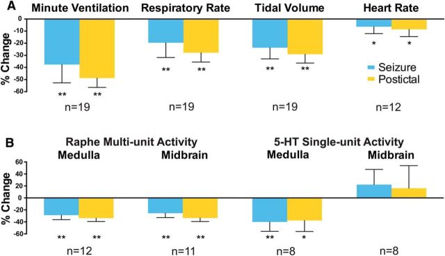
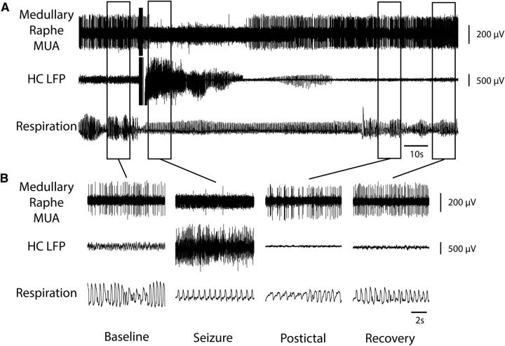
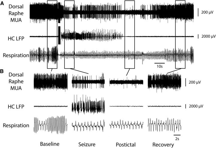
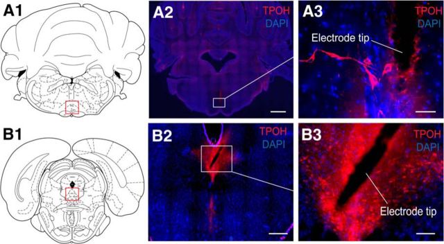
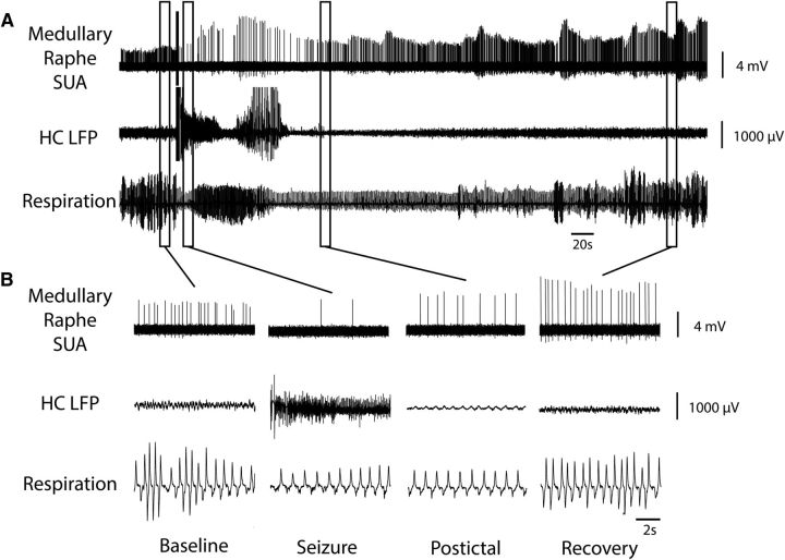
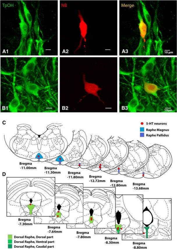
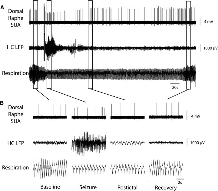
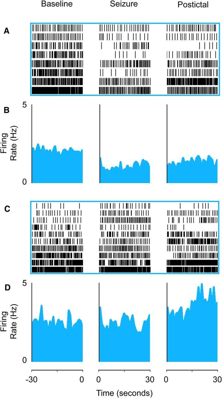
Impaired breathing, cardiac function, and arousal during and after seizures are important causes of morbidity and mortality. Previous work suggests that these changes are associated with depressed brainstem function in the ictal and post-ictal periods. Lower brainstem serotonergic systems are postulated to play an important role in cardiorespiratory changes during and after seizures, whereas upper brainstem serotonergic and other systems regulate arousal. However, direct demonstration of seizure-associated neuronal activity changes in brainstem serotonergic regions has been lacking. Here, we performed multiunit and single-unit recordings from medullary raphe and midbrain dorsal raphe nuclei in an established rat seizure model while measuring changes in breathing rate and depth as well as heart rate. Serotonergic neurons were identified by immunohistochemistry. Respiratory rate, tidal volume, and minute ventilation were all significantly decreased during and after seizures in this model. We found that population firing of neurons in the medullary and midbrain raphe on multiunit recordings was significantly decreased during the ictal and post-ictal periods. Single-unit recordings from identified serotonergic neurons in the medullary raphe revealed highly consistently decreased firing during and after seizures. In contrast, firing of midbrain raphe serotonergic neurons was more variable, with a mixture of increases and decreases. The markedly suppressed firing of medullary serotonergic neurons supports their possible role in simultaneously impaired cardiorespiratory function in seizures. Decreased arousal likely arises from depressed population activity of several neuronal pools in the upper brainstem and forebrain. These findings have important implications for preventing morbidity and mortality in people living with epilepsy.

Significance statement: Seizures often cause impaired breathing, cardiac dysfunction, and loss of consciousness. The brainstem and, specifically, brainstem serotonin neurons are thought to play an important role in controlling breathing, cardiac function, and arousal. We used an established rat seizure model to study the overall neuronal activity in the brainstem as well as firing of specific serotonin neurons while measuring cardiorespiratory function. Our results demonstrated overall decreases in brainstem neuronal activity and marked downregulation of lower brainstem serotonin neuronal firing in association with decreased breathing and heart rate during and after seizures. These findings point the way toward new treatments to augment brainstem function and serotonin, aiming to prevent seizure complications and reduce morbidity and mortality in people living with epilepsy.

Keywords: SUDEP; brainstem; consciousness; respiratory; serotonin; temporal lobe epilepsy.


Figures

Figure 1.
Figure 1.
Effects of seizures on cardiorespiratory function and brainstem neuronal activity. A, Cardiorespiratory effects of seizures induced by 2 s of hippocampal stimulation. Mean minute ventilation, respiratory rate, tidal volume, and heart rate all decrease in the ictal and post-ictal periods compared with 30 s of preseizure baseline. B, Effects of seizures on neuronal activity in the medullary raphe and midbrain dorsal raphe nuclei. Mean MUA from the medullary and midbrain raphe and SUA from medullary 5-HT neurons decreased in the ictal and post-ictal periods. Mean SUA of 5-HT neurons in the midbrain dorsal raphe was not significantly changed by seizures. Locations of recording electrodes and neurons are shown in Figures 4 and 6. Sample sizes (n, animals) are indicated. One seizure per animal was analyzed for A. For B, the number of seizures analyzed were 63 for MUA in medulla, 58 for MUA in midbrain, 8 for SUA in medulla, and 10 for SUA in midbrain. Error bars indicate SEM. *p ≤ 0.05; **p < 0.005, Holm–Bonferroni corrected.
Figure 2.
Figure 2.
Decreased MUA in medullary raphe and reduced breathing during seizure. A, Seizure induced by 2 s stimulation of HC. After the stimulus, fast polyspike activity is seen in the HC LFP. MUA from the medullary raphe shows marked suppression of neuronal firing during the seizure with gradual recovery in the post-ictal period. Respiratory trace shows breathing flow amplitude (proportional to volume) and rate both decreased markedly during the ictal and post-ictal periods. B, Expanded segments of data from baseline, seizure, post-ictal, and recovery periods from the boxed regions in A.
Figure 3.
Figure 3.
Decreased MUA in midbrain dorsal raphe and reduced breathing during seizure. A, Seizure induced by 2 s of stimulation of HC. After the stimulus, fast polyspike activity is seen in the HC LFP. MUA from the midbrain dorsal raphe shows marked suppression of neuronal firing during the seizure with gradual recovery in the post-ictal period. Respiratory trace shows breathing flow amplitude (proportional to volume) and rate both decreased markedly during the ictal and post-ictal periods. B, Expanded segments of data from baseline, seizure, post-ictal, and recovery periods from the boxed regions in A.
Figure 4.
Figure 4.
Serotonergic neurons in regions of medullary and midbrain MUA recordings. A, Histology example from medullary raphe MUA recording: a coronal section at approximately AP −10.3 mm. B, Example from midbrain dorsal raphe MUA recording: coronal section at approximately −7.64 mm. Red boxes in A1 and B1 represent the general regions where serotonergic nuclei are located in the medulla and midbrain, respectively. A2 and B2 show an overview of tryptophan hydroxylase (TPOH) and nuclear DAPI staining of the target slices. A3 and B3 are expanded views of white boxes in A2 and B2, showing serotonergic and other neurons in the vicinity of the MUA electrode tips. Scale bars: A2, 1 mm; B2, 500 μm; A3, 50 μm; B3, 100 μm. A1 and A2 are reproduced with permission from Paxinos and Watson (1998).
Figure 5.
Figure 5.
Medullary raphe 5-HT neuron decreases firing during and after seizure. A, Seizure induced by 2 s stimulation of HC. After the stimulus, fast polyspike activity is seen in the HC LFP. Single unit activity (SUA) from the medullary raphe serotonergic neuron shows marked suppression of neuronal firing during the seizure with gradual recovery in the post-ictal period. Respiratory trace shows breathing flow amplitude and rate both decreased markedly during the ictal and post-ictal periods. B, Expanded segments of data from baseline, seizure, post-ictal, and recovery periods from the boxed regions in A.
Figure 6.
Figure 6.
Histology of 5-HT neurons studied by juxtacellular recordings in medulla and midbrain. A, Labeled medullary raphe 5-HT cell recorded juxtacellularly, stained for tryptophan hydroxylase (TPOH) in A1, Neurobiotin (NBiotin) in A2, and merge in A3. B, Labeled midbrain dorsal raphe 5-HT cell recorded juxtacellularly, stained for TPOH in B1, Neurobiotin in B2, and merge in B3. Scale bars, 10 μm. C, Locations of all recorded 5-HT neurons in the medulla (n = 8). Identified 5-HT neurons were located in regions of raphe magnus and raphe pallidus. D, Locations of all recorded 5-HT neurons in the midbrain (n = 10). Identified 5-HT neurons were located in the following regions of the dorsal raphe dorsal, ventral, and caudal subnuclei. Coronal section schematics reproduced with permission from Paxinos and Watson (1998).
Figure 7.
Figure 7.
Midbrain raphe 5-HT neuron does not decrease firing during seizure. A, Seizure induced by 2 s stimulation of HC. After the stimulus, fast polyspike activity is seen in the HC LFP. Single-unit activity (SUA) from the midbrain dorsal raphe serotonergic neuron continues firing without major overall changes during or after the seizure. Respiratory trace again shows breathing flow amplitude and rate both decreased markedly during the ictal and post-ictal periods. B, Expanded segments of data from baseline, seizure, post-ictal, and recovery periods from the boxed regions in A.
Figure 8.
Figure 8.
Serotonergic neurons in medulla decrease firing during seizures, whereas firing of midbrain serotonin neurons is mixed. A, B, Medullary raphe serotonergic neurons showed decreased firing during seizures and in the post-ictal period. A, Raster plot of recovered 5-HT medullary raphe cells (n = 8 cells from 8 animals). B, Mean firing rate histogram data. C, D, Midbrain raphe serotonergic neurons had variable changes in firing rate during seizures. C, Raster plot of recovered 5-HT midbrain raphe neurons in dorsal raphe (n = 10 cells from 8 animals). D, Mean firing rate histogram data. Neurons in raster plots (A, C) are ordered based on baseline firing rate for visualization. Histogram data (B, D) are presented as curvilinear plots with firing rates in 1 s nonoverlapping bins. Data are from the 30 s baseline immediately before seizures, the first 30 s after seizure onset, and the first 30 s after seizure end in the post-ictal period. Anatomical locations of all recorded neurons are shown in Figure 7.

References

    1. Bateman LM, Spitz M, Seyal M. Ictal hypoventilation contributes to cardiac arrhythmia and SUDEP: report on two deaths in video-EEG-monitored patients. Epilepsia. 2010;51:916–920. doi: 10.1111/j.1528-1167.2009.02513.x. - DOI - PubMed
    1. Blessing WW. The lower brainstem and bodily homeostasis. Oxford: OUP; 1997.
    1. Blumenfeld H. Impaired consciousness in epilepsy. Lancet Neurol. 2012;11:814–826. doi: 10.1016/S1474-4422(12)70188-6. - DOI - PMC - PubMed
    1. Blumenfeld H, Rivera M, McNally KA, Davis K, Spencer DD, Spencer SS. Ictal neocortical slowing in temporal lobe epilepsy. Neurology. 2004a;63:1015–1021. doi: 10.1212/01.WNL.0000141086.91077.CD. - DOI - PubMed
    1. Blumenfeld H, McNally KA, Vanderhill SD, Paige AL, Chung R, Davis K, Norden AD, Stokking R, Studholme C, Novotny EJ, Jr, Zubal IG, Spencer SS. Positive and negative network correlations in temporal lobe epilepsy. Cereb Cortex. 2004b;14:892–902. doi: 10.1093/cercor/bhh048. - DOI - PubMed

Publication types

LinkOut - more resources