. 2016 Apr 29;90(10):4926-38.
doi: 10.1128/JVI.02517-15. Print 2016 May 15.

The Human Cytomegalovirus UL116 Gene Encodes an Envelope Glycoprotein Forming a Complex with gH Independently from gL

Affiliations

The Human Cytomegalovirus UL116 Gene Encodes an Envelope Glycoprotein Forming a Complex with gH Independently from gL

Stefano Caló et al. J Virol. .

Abstract

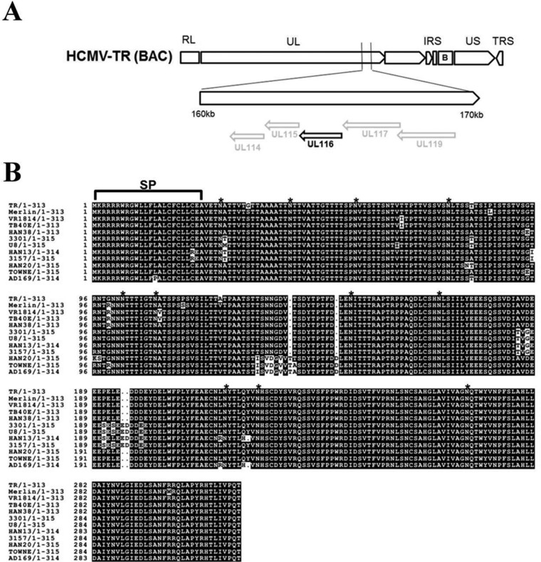
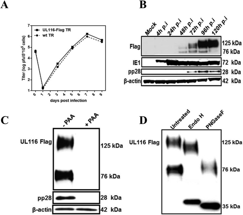
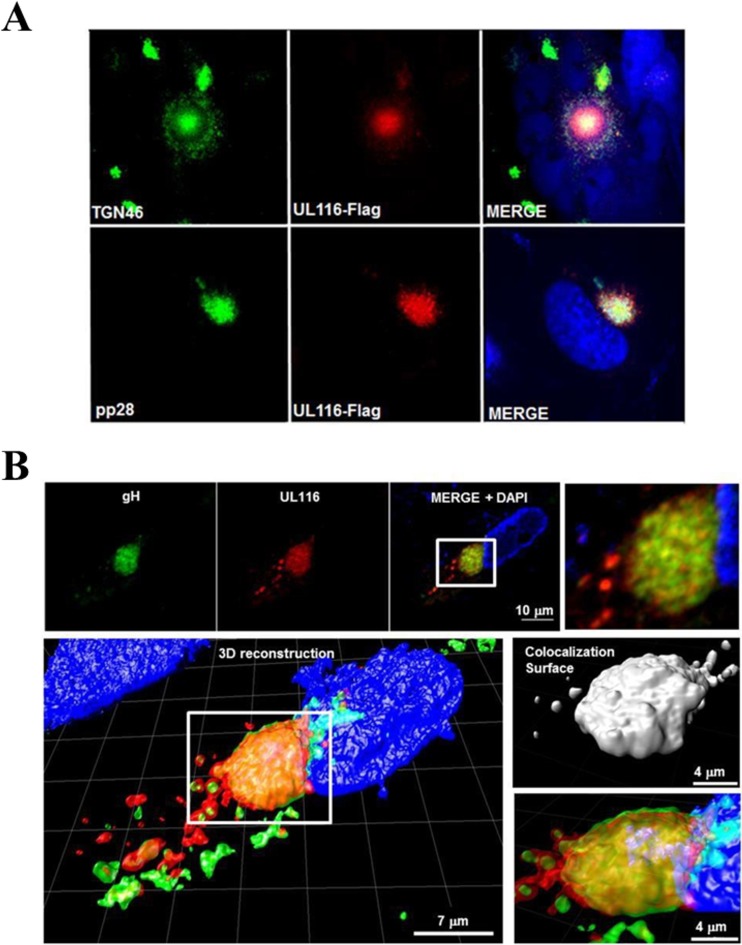
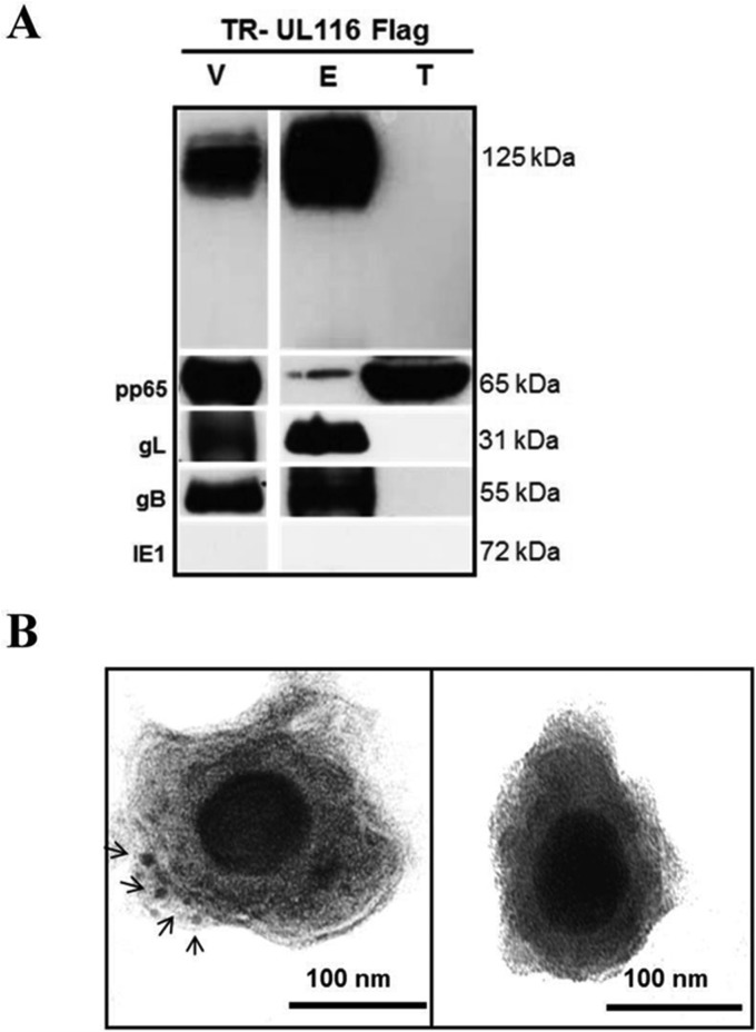
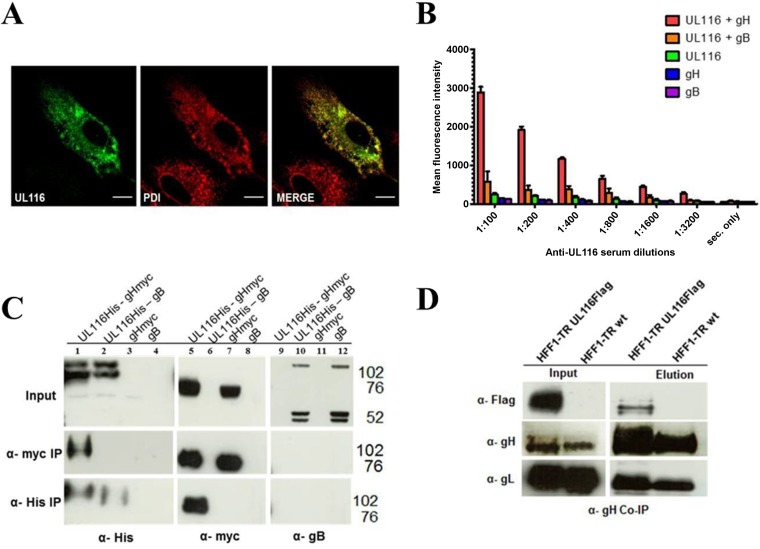
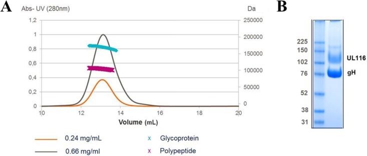
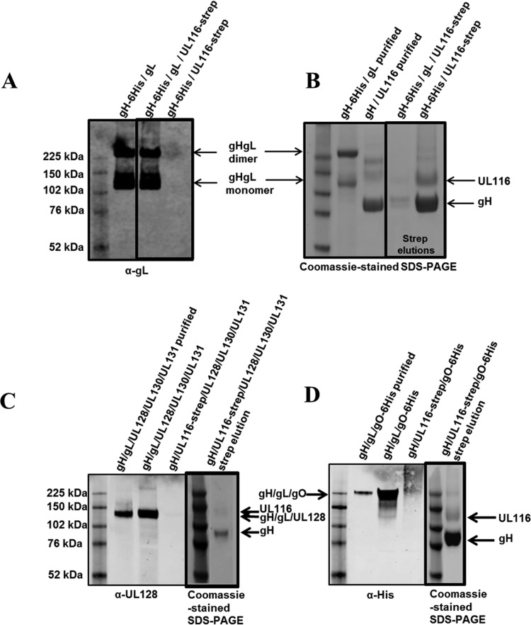
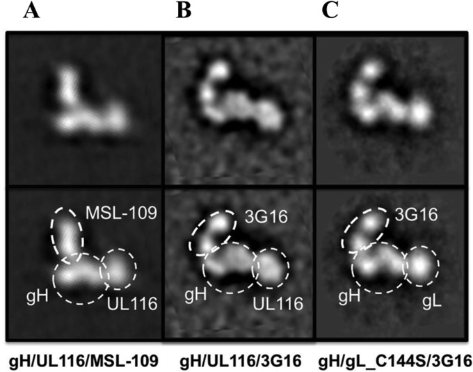
Human cytomegalovirus (HCMV) is a major cause of morbidity and mortality in transplant patients and is the leading viral cause of birth defects after congenital infection. HCMV infection relies on the recognition of cell-specific receptors by one of the viral envelope glycoprotein complexes. Either the gH/gL/gO or the gH/gL/UL128/UL130/UL131A (Pentamer) complex has been found to fulfill this role, accounting for HCMV entry into almost all cell types. We have studied the UL116 gene product, a putative open reading frame identified by in silico analysis and predicted to code for a secreted protein. Virus infection experiments in mammalian cells demonstrated that UL116 is expressed late in the HCMV replication cycle and is a heavily glycosylated protein that first localizes to the cellular site of virus assembly and then inserts into the virion envelope. Transient-transfection studies revealed that UL116 is efficiently transported to the plasma membrane when coexpressed with gH and that gL competes with UL116 for gH binding. Further evidence for gH/UL116 complex formation was obtained by coimmunoprecipitation experiments on both transfected and infected cells and biochemical characterization of the purified complex. In summary, our results show that the product of the UL116 gene is an HCMV envelope glycoprotein that forms a novel gH-based complex alternative to gH/gL. Remarkably, the gH/UL116 complex is the first herpesvirus gH-based gL-less complex.

Importance: HCMV infection can cause severe disease in immunocompromised adults and infants infected in utero The dissection of the HCMV entry machinery is important to understand the mechanism of viral infection and to identify new vaccine antigens. The gH/gL/gO and gH/gL/UL128/UL130/UL131 (Pentamer) complexes play a key role in HCMV cell entry and tropism. Both complexes are formed by an invariant gH/gL scaffold on which the other subunits assemble. Here, we show that the UL116 gene product is expressed in infected cells and forms a heterodimer with gH. The gH/UL116 complex is carried on the infectious virions, although in smaller amounts than gH/gL complexes. No gH/UL116/gL ternary complex formed in transfected cells, suggesting that the gH/UL116 complex is independent from gL. This new gH-based gL-free complex represents a potential target for a protective HCMV vaccine and opens new perspectives on the comprehension of the HCMV cell entry mechanism and tropism.


Figures

FIG 1
FIG 1
UL116 position in the HCMV TR genome and sequence conservation among laboratory-adapted and clinical HCMV strains. (A) ORF map of the TR BAC clone used in this work. Arrows indicate the relative orientations of the repeated and unique ORF blocks. The UL116 gene, in boldface, is located between UL115 (gL) and UL117 genes on the antisense strand. (B) T-Coffee primary amino acid sequence multialignment showing 98% UL116 gene conservation among members of a consistent pool of HCMV strains. Asterisks indicate the predicted 14 N-glycosylation sites. SP indicates the N-terminal predicted signal peptide. RL, repeat long; UL, unique long; IRS, internal repeat short; US, unique short; TRS, terminal repeat short; B, BAC inserts.
FIG 2
FIG 2
UL116 expression kinetics and carbohydrate addition in HCMV-infected fibroblasts. (A) Multiple-step growth curve analysis of the reconstituted virus UL116-Flag TR and of the parental HCMV strain TR. Time point 0 titers represent input inocula, and each data point represents averages from three independent wells. (B) Uninfected (mock) and TR-UL116-Flag-infected HFF-1 cells at an multiplicity of infection of 5 were harvested at the indicated times postinfection. Equivalent amounts of cell lysates were subjected to SDS-PAGE under reducing conditions and analyzed by immunoblotting with anti-Flag, HCMV IE1, and pp28 protein antibodies as indicated on the left. Actin detection was used as a protein loading control. (C) TR-UL116-Flag infections of HFF cells were performed in the presence (right) or absence (left) of phosphonoacetic acid (PAA), an inhibitor of HCMV late-phase protein expression. Five days postinfection, lysates were prepared from infected cells, and UL116 and pp28 expression was detected by Western blotting using anti-Flag and anti-pp28 antibodies, respectively. (D) Equal amounts of cell lysates of TR UL116-Flag-infected HFF-1 cells (72 h p.i.) were subjected to glycosidase treatments: left lane, untreated control; middle and right lanes, endoglycosidase H (Endo H) and PNGase F treatments, respectively. Proteins were separated on SDS-PAGE under reducing conditions, and UL116 was detected by immunoblot analysis using an anti-Flag antibody.
FIG 3
FIG 3
Intracellular localization of UL116 in infected HFF-1 cells. (A) HFF-1 cells were infected with the recombinant virus TR-UL116-Flag at an MOI of 5 for 72 h. Cells then were fixed, permeabilized, and stained. UL116 detection was achieved by anti-Flag (in red) in combination with either the cellular marker TGN 46 (upper) or the HCMV tegument protein pp28 (lower). Cell nuclei are stained blue. The merge panels show colocalization of the red and green signals. (B) Same experiment as that described for panel A, except that mouse anti-gH was used. A 3D reconstruction and the colocalization surface of the UL116 and gH signals are shown in the lower images.
FIG 4
FIG 4
Localization of UL116 on the virion envelope. (A) Western blotting was performed on purified virions from virus expressing Flag-tagged UL116. The virion total lysate (V), envelope fraction (E), and tegument/capsid fraction (T) were probed for the indicated antigens. (B) Immunogold EM staining for anti-Flag MAb of purified TR-UL116Flag and TR wild type (left and right, respectively). Black arrows indicate gold particles. Original magnification, ×40,000.
FIG 5
FIG 5
UL116 interaction with gH in HEK293T transfected cells. (A) ARPE-19 cells transiently expressing UL116-myc were fixed, permeabilized, and stained with anti-myc (green) and anti-PDI (red) specific antibodies prior to confocal immunofluorescence microscopy. Scale bars represent 10 μm. (B) Detection of UL116 by fluorescence-activated cell sorter (FACS) analysis on nonpermeabilized HEK293T cells. Cells were transfected with expression vectors for UL116 as well as gH and gB, both alone and in combination, as indicated. Forty-eight hours posttransfection, cells were stained at 4°C with anti-UL116 polyclonal mouse serum at different dilutions. Excess probe was removed by washing in PBS, and then cells were fixed and stained with Alexa Fluor 488-conjugated anti-mouse antibody. For each point, 10,000 cells were analyzed and the mean fluorescent intensity of Alexa Fluor 488-positive cells was reported. Secondary antibody only was used as a negative control. Experiments were performed in triplicate. (C) UL116-gH coimmunoprecipitation. Lysates from HEK293T cells transiently expressing UL116-his/gH-myc, UL116-his, gB, and UL116-his/gB were subjected to parallel immunoprecipitation (IP) experiments (antibodies used are specified on the left) with both covalently linked magnetic anti-His and anti-myc beads. Total lysates (input) and eluted samples were separated by SDS-PAGE and analyzed by immunoblotting for both the His and myc tag (indicated at the bottom). (D) Coimmunoprecipitation (Co-IP) of the UL116-gH complex in infected cells. Cell lysates were prepared from HFF-1 infected with HCMV-TR UL116 Flag and wt TR separately (5 dpi). Complexes were captured using the human monoclonal antibody MSL-109. Elutions and crude extracts were subjected to immunoblotting using a rabbit anti-gH polyclonal serum, an anti-Flag M2 clone monoclonal MAb, and a rabbit polyclonal serum specific for gL.
FIG 6
FIG 6
MALS analysis of purified gH/ULL116 complex. (A) SEC profile of the gH/UL116 complex at two different concentrations (gray and orange traces) and molecular mass estimates for the polypeptide only (110 kDa) and for the glycosylated complex (160 kDa) determined by MALS (pink and cyan lines, respectively). The left y axis pertains to UV absorbance (Abs), while the right y axis represents the molecular weight. (B) SDS-PAGE analysis under nonreducing conditions of the gH/UL116 complex used in panel A.
FIG 7
FIG 7
Western blotting and SDS-PAGE for gH/gL, gH/gL/UL116, and gH/UL116 transfections. (A) Anti-gL Western blotting (nonreducing) on conditioned media from Expi293 cells transiently transfected with DNA encoding the indicated proteins. (B) gH-6His/UL116-strep and gH-6His/gL/UL116-strep samples were Strep-tag affinity purified from the medium, and the elution fraction was run in SDS-PAGE. Coomassie-stained nonreducing SDS-PAGE gels show purified gH/gL and gH/UL116 (controls) followed by strep elution samples. (C and D) Anti-UL128 (C) and anti-His (D) Western blotting (nonreducing) on purified protein alongside conditioned medium from Expi293 cells transiently transfected with the DNA encoding the indicated proteins. Samples containing UL116-strep then were Strep-tag affinity purified from the medium, and the elution fractions were run in SDS-PAGE. Coomassie-stained nonreducing SDS-PAGE gel showing elution fractions are in thickly lined boxes in panels C and D.
FIG 8
FIG 8
Single-particle EM of the gH/UL116 complex. Purified gH/UL116/MSL-109 (A), gH/UL116/3G16 (B), and gH/gL_C144S/3G16 (C) ternary complexes were analyzed by single-particle electron microscopy. Individual particles, representing a specific 2D view of the complex, were aligned and used to generate 2D class averages. Comparison among the class averages of these 3 complexes and also with previous ones obtained for gH/gL and gH/gL/gO bound to MSL-109 or 3G16 (72) suggest the position of individual subunits and Fabs.

References

    1. Mocarski JE, Shenk T, Griffiths PD, Pass RF. 2013. Cytomegaloviruses, p 1960–2014. In Knipe DM, Howley PM, Cohen JI, Griffin DE, Lamb RA, Martin MA, Racaniello VR, Roizman B (ed), Fields virology, 6th ed Lippincott Williams & Wilkins, Philadelphia, PA.
    1. Cannon MJ. 2009. Congenital cytomegalovirus (CMV) epidemiology and awareness. J Clin Virol 46(Suppl 4):S6–S10. doi:10.1016/j.jcv.2009.09.002. - DOI - PubMed
    1. Britt W. 2008. Manifestations of human cytomegalovirus infection: proposed mechanisms of acute and chronic disease. Curr Topics Microbiol Immunol 325:417–470. - PubMed
    1. Krause PR, Bialek SR, Boppana SB, Griffiths PD, Laughlin CA, Ljungman P, Mocarski ES, Pass RF, Read JS, Schleiss MR, Plotkin SA. 2013. Priorities for CMV vaccine development. Vaccine 32:4–10. doi:10.1016/j.vaccine.2013.09.042. - DOI - PMC - PubMed
    1. Stratton KR, Durch JS, Lawrence RS. 2001. Vaccines for the 21st century: a tool for a decisionmaking. National Academy Press, Washington, DC. - PubMed

Publication types

Substances

LinkOut - more resources