. 2016 Apr 14;90(9):4780-4795.
doi: 10.1128/JVI.02586-15. Print 2016 May.

MicroRNA-19b-3p Modulates Japanese Encephalitis Virus-Mediated Inflammation via Targeting RNF11

Affiliations

MicroRNA-19b-3p Modulates Japanese Encephalitis Virus-Mediated Inflammation via Targeting RNF11

Usama Ashraf et al. J Virol. .

Abstract

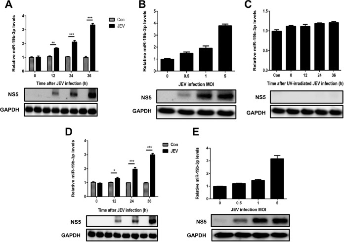
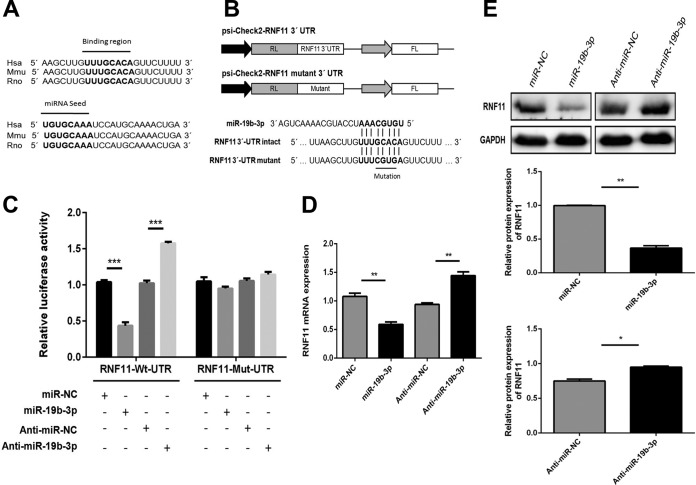
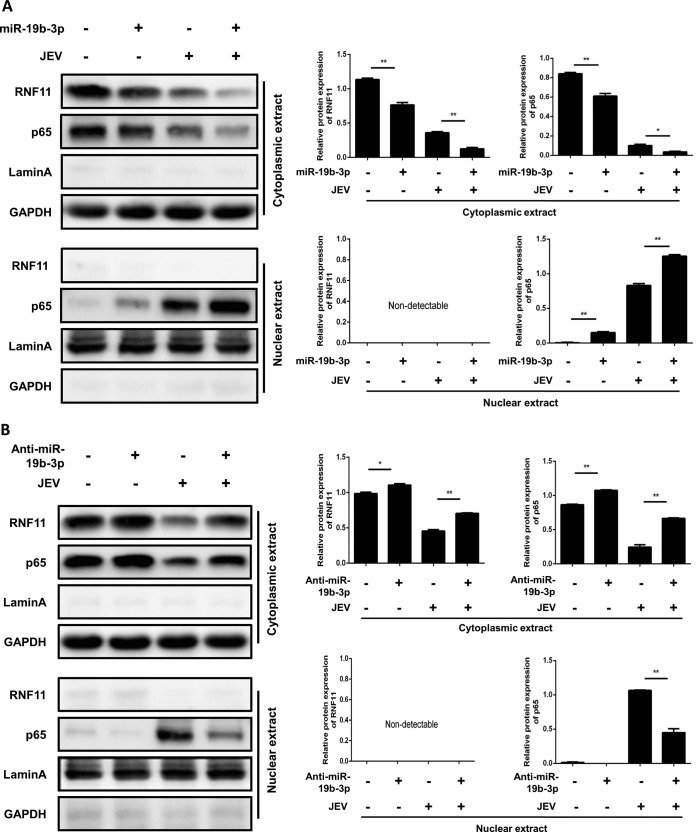
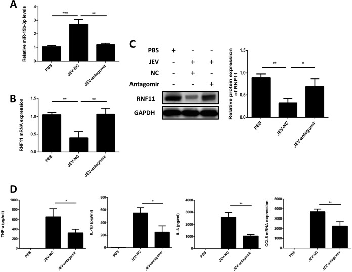
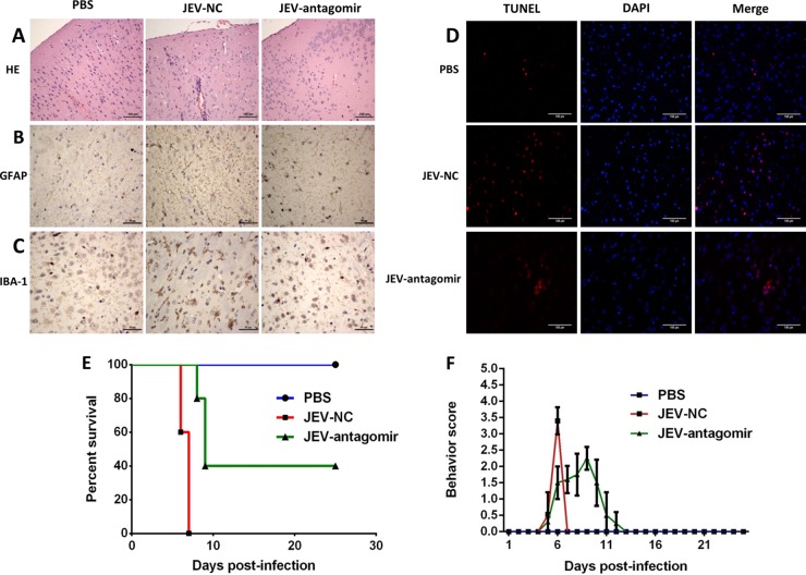
Japanese encephalitis virus (JEV) can invade the central nervous system and consequently induce neuroinflammation, which is characterized by profound neuronal cell damage accompanied by astrogliosis and microgliosis. Albeit microRNAs (miRNAs) have emerged as major regulatory noncoding RNAs with profound effects on inflammatory response, it is unknown how astrocytic miRNAs regulate JEV-induced inflammation. Here, we found the involvement of miR-19b-3p in regulating the JEV-induced inflammatory responsein vitroandin vivo The data demonstrated that miR-19b-3p is upregulated in cultured cells and mouse brain tissues during JEV infection. Overexpression of miR-19b-3p led to increased production of inflammatory cytokines, including tumor necrosis factor alpha, interleukin-6, interleukin-1β, and chemokine (C-C motif) ligand 5, after JEV infection, whereas knockdown of miR-19b-3p had completely opposite effects. Mechanistically, miR-19b-3p modulated the JEV-induced inflammatory response via targeting ring finger protein 11, a negative regulator of nuclear factor kappa B signaling. We also found that inhibition of ring finger protein 11 by miR-19b-3p resulted in accumulation of nuclear factor kappa B in the nucleus, which in turn led to higher production of inflammatory cytokines.In vivosilencing of miR-19b-3p by a specific antagomir reinvigorates the expression level of RNF11, which in turn reduces the production of inflammatory cytokines, abrogates gliosis and neuronal cell death, and eventually improves the survival rate in the mouse model. Collectively, our results demonstrate that miR-19b-3p positively regulates the JEV-induced inflammatory response. Thus, miR-19b-3p targeting may constitute a thought-provoking approach to rein in JEV-induced inflammation.

Importance: Japanese encephalitis virus (JEV) is one of the major causes of acute encephalitis in humans worldwide. The pathological features of JEV-induced encephalitis are inflammatory reactions and neurological diseases resulting from glia activation. MicroRNAs (miRNAs) are small noncoding RNAs that regulate gene expression posttranscriptionally. Accumulating data indicate that miRNAs regulate a variety of cellular processes, including the host inflammatory response under pathological conditions. Recently, a few studies demonstrated the role of miRNAs in a JEV-induced inflammatory response in microglia; however, their role in an astrocyte-derived inflammatory response is largely unknown. The present study reveals that miR-19b-3p targets ring finger protein 11 in glia and promotes inflammatory cytokine production by enhancing nuclear factor kappa B activity in these cells. Moreover, administration of an miR-19b-3p-specific antagomir in JEV-infected mice reduces neuroinflammation and lethality. These findings suggest a new insight into the molecular mechanism of the JEV-induced inflammatory response and provide a possible therapeutic entry point for treating viral encephalitis.


Figures

FIG 1
FIG 1
miR-19b-3p expression is upregulated after JEV infection. (A and B) U251 cells were infected with JEV at an MOI of 5 for the indicated times (A) or at the indicated MOIs for 36 h (B). (C) U251 cells were incubated with UV-inactivated JEV at an MOI of 5 for 36 h. (D and E) BV2 cells were infected with JEV at an MOI of 5 for the indicated times (D) or at the indicated MOIs for 36 h (E). The levels of miR-19b-3p were detected by quantitative real-time PCR (upper panels). Western blotting was performed to examine the expression of JEV NS5 protein (lower panels). GAPDH expression was verified as a loading control. All data are representative of at least three independent experiments. *, P < 0.05; **, P < 0.01; ***, P < 0.001. Con, control.
FIG 2
FIG 2
miR-19b-3p enhances JEV-mediated production of inflammatory cytokines. (A) U251 cells were transfected with miR-19b-3p mimics or control oligonucleotides (final concentration, 50 nM) for 24 h and then either left uninfected or infected with JEV at an MOI of 5 for 36 h. The level of miR-19b-3p was analyzed by quantitative real-time PCR and normalized to the U6 level. ***, P < 0.001. (B) The protein levels of TNF-α, IL-6, and IL-1β were analyzed by ELISA. Data represent means ± SD from three independent experiments performed in duplicate. *, P < 0.05; **, P < 0.01. CCL5 mRNA levels were determined by quantitative real-time PCR and normalized to the expression of β-actin in each sample. Data represent means ± SD from three independent experiments. *, P < 0.05; ***, P < 0.001. (C) The transfected U251 cells were infected with JEV at an MOI of 5. Cells were collected at the indicated time points, and titers of infectious virus in the culture supernatants were determined by plaque assay. The data represent three independent experiments with identical results. (D) BV2 cells were transfected with miR-19b-3p mimics or control oligonucleotides (final concentration, 50 nM) for 24 h and then either left uninfected or infected with JEV at an MOI of 5 for 36 h. The level of miR-19b-3p was analyzed by quantitative real-time PCR and normalized to the U6 level. ***, P < 0.001. (E) The protein levels of TNF-α, IL-6, and IL-1β were analyzed by ELISA. Data represent means ± SD from three independent experiments performed in duplicate. *, P < 0.05; **, P < 0.01. CCL5 mRNA levels were determined by quantitative real-time PCR and normalized to the expression of β-actin in each sample. Data represent means ± SD from three independent experiments. **, P < 0.01.
FIG 3
FIG 3
Inhibition of miR-19b-3p suppresses JEV-mediated production of inflammatory cytokines. (A) U251 cells were transfected with miR-19b-3p inhibitors or control oligonucleotides (final concentration, 50 nM) for 24 h and then either left uninfected or infected with JEV at an MOI of 5 for 36 h. The level of miR-19b-3p was analyzed by quantitative real-time PCR and normalized to the U6 level. **, P < 0.01; ***, P < 0.001. (B) The protein levels of TNF-α, IL-6, and IL-1β were analyzed by ELISA. Data represent means ± SD from three independent experiments performed in duplicate. *, P < 0.05; **, P < 0.01. CCL5 mRNA levels were determined by quantitative real-time PCR and normalized to the expression of β-actin in each sample. Data represent means ± SD from three independent experiments. *, P < 0.05; ***, P < 0.001. (C) The transfected U251 cells were infected with JEV at an MOI of 5. Cells were collected at the indicated time points, and titers of infectious virus in the culture supernatants were determined by plaque assay. The data represent three independent experiments with identical results. (D) BV2 cells were transfected with miR-19b-3p inhibitors or control oligonucleotides (final concentration, 50 nM) for 24 h and then either left uninfected or infected with JEV at an MOI of 5 for 36 h. The level of miR-19b-3p was analyzed by quantitative real-time PCR and normalized to the U6 level. **, P < 0.01. (E) The protein levels of TNF-α, IL-6, and IL-1β were analyzed by ELISA. Data represent means ± SD from three independent experiments performed in duplicate. *, P < 0.05. CCL5 mRNA levels were determined by quantitative real-time PCR and normalized to the expression of β-actin in each sample. Data represent means ± SD from three independent experiments. *, P < 0.05; **, P < 0.01.
FIG 4
FIG 4
miR-19b-3p directly targets RNF11. (A) Conservation of the miR-19b-3p target sequence in RNF11 among different species (upper panel), and conservation of the sequence of miR-19b-3p among different species (lower panel). Hsa, Homo sapiens (human); Mmu, Mus musculus (mouse); Rno; Rattus norvegicus (rat). (B) Schematic diagram showing dual-luciferase reporter constructs harboring the 3′ UTR of RNF11 with putative miR-19b-3p binding site. The lower panel shows the alignment of miR-19b-3p and its target site in the 3′ UTR of RNF11. Four mutated nucleotides of the target site are underlined. RL, Renilla luciferase; FL, firefly luciferase. (C) U251 cells were cotransfected with miR-19b-3p mimics, miR-19b-3p inhibitors, or the corresponding control oligonucleotide (final concentration, 50 nM) together with a wild-type or mutated RNF11 3′ UTR dual-luciferase reporter plasmid, and Renilla luciferase activity was measured and normalized to firefly luciferase activity after 24 h. (D and E) U251 cells were transfected with miR-19b-3p mimics, miR-19b-3p inhibitors, or the corresponding control oligonucleotide (final concentration, 50 nM), and then RNF11 mRNA (C) and protein levels (D) were determined after 24 h by quantitative real-time PCR and immunoblotting, respectively. Data represent means ± SD from three independent experiments. *, P < 0.05; **, P < 0.01; ***, P < 0.001. Protein levels were quantified with immunoblot scanning and normalized to the amount of GAPDH expression.
FIG 5
FIG 5
RNF11 expression is downregulated after JEV infection. (A) U251 cells were infected with JEV at an MOI of 5 for the indicated times, and then RNF11 mRNA expression levels were determined by quantitative real-time PCR. (B) U251 cells were infected with JEV at an MOI of 5 for the indicated times, and then RNF11 protein levels were determined with immunoblotting. (C and D) BV2 cells were infected with JEV at an MOI of 5 for the indicated times, and then RNF11 mRNA (C) and protein (D) expression levels were determined by quantitative real-time PCR and immunoblotting, respectively. Data represent means ± SD from three independent experiments. *, P < 0.05; **, P < 0.01; ***, P < 0.001. Protein levels were quantified with immunoblot scanning and normalized to the amount of GAPDH expression.
FIG 6
FIG 6
Regulation of JEV-induced production of inflammatory cytokines by miR-19b-3p is achieved through RNF11. (A) U251 cells were transfected with siRNF11 or nonspecific control siRNA (siNC) (final concentration, 50 nM) for 24 h, and then RNF11 protein levels were measured by immunoblotting. Protein levels were quantified by immunoblot scanning and normalized to the amount of GAPDH expression. (B) U251 cells were cotransfected with miR-19b-3p inhibitors or control oligonucleotides and siRNF11 or a nonspecific control siRNA (final concentration, 50 nM) for 24 h and then infected with JEV at an MOI of 5 for 36 h. The levels of miR-19b-3p were analyzed by quantitative real-time PCR and normalized to the U6 level. **, P < 0.01; ***, P < 0.001. (C) The protein levels of TNF-α, IL-6, and IL-1β were analyzed by ELISA. Data represent means ± SD from three independent experiments performed in duplicate. *, P < 0.05; **, P < 0.01; ***, P < 0.001. CCL5 mRNA levels were determined with quantitative real-time PCR and normalized to the expression of β-actin in each sample. Data represent means ± SD from three independent experiments. *, P < 0.05; **, P < 0.01; ***, P < 0.001.
FIG 7
FIG 7
miR-19b-3p activates the NF-κB pathway in JEV-infected astrocytes. (A and B) U251 cells were transfected with miR-19b-3p mimics, inhibitors, or their control oligonucleotides (final concentration, 50 nM) for 24 h and then either left uninfected or infected with JEV at an MOI of 5 for 36 h. The cytosolic extracts (upper panel) and nuclear extracts (lower panel) were isolated and subjected to immunoblotting with antibodies against RNF11, NF-κB p65, lamin A, and GAPDH. Lamin A was used as a marker for nuclei. GAPDH and lamin A were used as the loading controls. Protein levels were quantified by immunoblot scanning and normalized to the amount of GAPDH or lamin A expression. *, P < 0.05; **, P < 0.01.
FIG 8
FIG 8
miR-19b-3p activates NF-κB signaling via targeting RNF11 in JEV-infected astrocytes. U251 cells were cotransfected with miR-19b-3p inhibitors or control oligonucleotides and siRNF11 or a nonspecific control siRNA (final concentration, 50 nM) for 24 h and then infected with JEV at an MOI of 5 for 36 h. The cytosolic extracts (upper panel) and nuclear extracts (lower panel) were isolated and subjected to immunoblotting with antibodies against RNF11, NF-κB p65, lamin A, and GAPDH. Lamin A was used as a marker for nuclei. GAPDH and lamin A were used as the loading controls. Protein levels were quantified by immunoblot scanning and normalized to the amount of GAPDH or lamin A expression. *, P < 0.05; **, P < 0.01; ***, P < 0.001.
FIG 9
FIG 9
Antagomir-19b-3p treatment stimulates RNF11 expression and reduces inflammatory cytokine production in a mouse model of JEV infection. Mice were treated with antagomir negative control or antagomir-19b-3p (60 mg/kg body weight) after JEV infection, and brain samples were collected at day 6 postinfection. PBS, samples collected from PBS-challenged mice; JEV-NC, samples collected from JEV-infected mice treated with antagomir negative control; JEV-antagomir, samples collected from JEV-infected mice treated with antagomir-19b-3p. (A) The increase of miR-19b-3p expression was abrogated in JEV-infected mice by administration of antagomir-19b-3p. (B and C) Suppression of RNF11 was reversed by antagomir-19b-3p treatment in JEV-infected mice. RNF11 mRNA (B) and protein (C) levels were determined by quantitative real-time PCR and immunoblotting, respectively. Protein levels were quantified by immunoblot scanning and normalized to the amount of GAPDH expression. (D) Antagomir-19b-3p treatment decreased the production of inflammatory cytokines. The protein levels of TNF-α, IL-6, and IL-1β were analyzed by ELISA. CCL5 mRNA levels were determined by quantitative real-time PCR and normalized to the expression of β-actin. The effects of antagomir-19b-3p and the antagomir negative control were compared using two-tailed Student's t tests. Similar results were obtained in three mice. *, P < 0.05; **, P < 0.01; ***, P < 0.001.
FIG 10
FIG 10
Antagomir-19b-3p treatment reduces neuroinflammation and improves the survival rate in JEV-infected mice. (A) Antagomir-19b-3p treatment ameliorates histopathological changes in JEV-infected mice. H&E staining of brain sections was performed to examine the pathological changes. Scale bar, 100 μm. (B and C) Antagomir-19b-3p treatment reduces astrocytosis and microgliosis. Sections of brain were analyzed by IHC staining. (B) Activation of astrocytes was detected by anti-GFAP antibody (B). Activation of microglia was detected by anti-IBA-1 antibody (C). Scale bar, 50 μm. (D) Antagomir-19b-3p treatment decreases neuronal cell damage. The apoptotic cells in the brain sections were stained using a TUNEL assay kit. Scale bar, 100 μm. The figures are a representative of three mice with similar results. (E) Antagomir-19b-3p treatment improves the survival rate. Survival of mice in each group was recorded for 25 days after JEV infection (100 PFU/mice) intracranially. Data were collected and are shown as Kaplan-Meier survival curves (n = 5 for each group). (F) Behavior score chart showing the gradual alleviation of suffering following JEV infection. Behavior scoring is as follows: 0, no restriction of movement, no frequent blinking, no body stiffening, no hind limb paralysis; 1, no restriction of movement, frequent blinking, no body stiffening, no hind limb paralysis; 2, restriction of movement, frequent blinking, no body stiffening, no hind limb paralysis; 3, restriction of movement, body stiffening, no hind limb paralysis; 4, restriction of movement, closed eyes, body stiffening, hind limb paralysis, body tremors.

Similar articles

Cited by

References

    1. Erlanger TE, Weiss S, Keiser J, Utzinger J, Wiedenmayer K. 2009. Past, present, and future of Japanese encephalitis. Emerg Infect Dis 15:1–7. doi:10.3201/eid1501.080311. - DOI - PMC - PubMed
    1. Yun SI, Lee YM. 2014. Japanese encephalitis: the virus and vaccines. Hum Vaccin Immunother 10:263–279. doi:10.4161/hv.26902. - DOI - PMC - PubMed
    1. Mackenzie JS. 2005. Emerging zoonotic encephalitis viruses: lessons from Southeast Asia and Oceania. J Neurovirol 11:434–440. doi:10.1080/13550280591002487. - DOI - PMC - PubMed
    1. Mackenzie JS, Gubler DJ, Petersen LR. 2004. Emerging flaviviruses: the spread and resurgence of Japanese encephalitis, West Nile and dengue viruses. Nat Med 10:S98–109. doi:10.1038/nm1144. - DOI - PubMed
    1. Campbell GL, Hills SL, Fischer M, Jacobson JA, Hoke CH, Hombach JM, Marfin AA, Solomon T, Tsai TF, Tsu VD, Ginsburg AS. 2011. Estimated global incidence of Japanese encephalitis: a systematic review. Bull World Health Organ 89:766–774. doi:10.2471/BLT.10.085233. - DOI - PMC - PubMed

Publication types

MeSH terms