

Derivation and Characterization of a CD4-Independent, Non-CD4-Tropic Simian Immunodeficiency Virus
- PMID: 26937037
- PMCID: PMC4859711
- DOI: 10.1128/JVI.02851-15
Derivation and Characterization of a CD4-Independent, Non-CD4-Tropic Simian Immunodeficiency Virus
Abstract
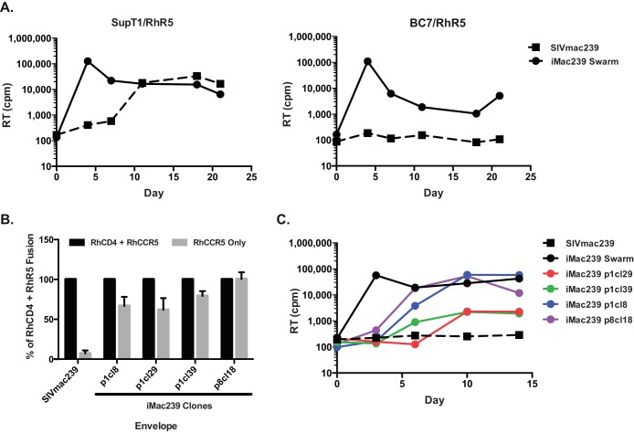
CD4 tropism is conserved among all primate lentiviruses and likely contributes to viral pathogenesis by targeting cells that are critical for adaptive antiviral immune responses. Although CD4-independent variants of human immunodeficiency virus (HIV) and simian immunodeficiency virus (SIV) have been described that can utilize the coreceptor CCR5 or CXCR4 in the absence of CD4, these viruses typically retain their CD4 binding sites and still can interact with CD4. We describe the derivation of a novel CD4-independent variant of pathogenic SIVmac239, termed iMac239, that was used to derive an infectious R5-tropic SIV lacking a CD4 binding site. Of the seven mutations that differentiate iMac239 from wild-type SIVmac239, a single change (D178G) in the V1/V2 region was sufficient to confer CD4 independence in cell-cell fusion assays, although other mutations were required for replication competence. Like other CD4-independent viruses, iMac239 was highly neutralization sensitive, although mutations were identified that could confer CD4-independent infection without increasing its neutralization sensitivity. Strikingly, iMac239 retained the ability to replicate in cell lines and primary cells even when its CD4 binding site had been ablated by deletion of a highly conserved aspartic acid at position 385, which, for HIV-1, plays a critical role in CD4 binding. iMac239, with and without the D385 deletion, exhibited an expanded host range in primary rhesus peripheral blood mononuclear cells that included CCR5(+) CD8(+) T cells. As the first non-CD4-tropic SIV, iMac239-ΔD385 will afford the opportunity to directly assess the in vivo role of CD4 targeting on pathogenesis and host immune responses.
Importance: CD4 tropism is an invariant feature of primate lentiviruses and likely plays a key role in pathogenesis by focusing viral infection onto cells that mediate adaptive immune responses and in protecting virions attached to cells from neutralizing antibodies. Although CD4-independent viruses are well described for HIV and SIV, these viruses characteristically retain their CD4 binding site and can engage CD4 if available. We derived a novel CD4-independent, CCR5-tropic variant of the pathogenic molecular clone SIVmac239, termed iMac239. The genetic determinants of iMac239's CD4 independence provide new insights into mechanisms that underlie this phenotype. This virus remained replication competent even after its CD4 binding site had been ablated by mutagenesis. As the first truly non-CD4-tropic SIV, lacking the capacity to interact with CD4, iMac239 will provide the unique opportunity to evaluate SIV pathogenesis and host immune responses in the absence of the immunomodulatory effects of CD4(+) T cell targeting and infection.
Copyright © 2016, American Society for Microbiology. All Rights Reserved.
Figures
Similar articles
-
Loss of a conserved N-linked glycosylation site in the simian immunodeficiency virus envelope glycoprotein V2 region enhances macrophage tropism by increasing CD4-independent cell-to-cell transmission.J Virol. 2014 May;88(9):5014-28. doi: 10.1128/JVI.02785-13. Epub 2014 Feb 19. J Virol. 2014. PMID: 24554659 Free PMC article.
-
CD4 independence of simian immunodeficiency virus Envs is associated with macrophage tropism, neutralization sensitivity, and attenuated pathogenicity.J Virol. 2002 Mar;76(6):2595-605. doi: 10.1128/jvi.76.6.2595-2605.2002. J Virol. 2002. PMID: 11861825 Free PMC article.
-
Induction of CD8+ cells able to suppress CCR5-tropic simian immunodeficiency virus SIVmac239 replication by controlled infection of CXCR4-tropic simian-human immunodeficiency virus in vaccinated rhesus macaques.J Virol. 2007 Nov;81(21):11640-9. doi: 10.1128/JVI.01475-07. Epub 2007 Aug 29. J Virol. 2007. PMID: 17728225 Free PMC article.
-
The SIV Envelope Glycoprotein, Viral Tropism, and Pathogenesis: Novel Insights from Nonhuman Primate Models of AIDS.Curr HIV Res. 2018;16(1):29-40. doi: 10.2174/1570162X15666171124123116. Curr HIV Res. 2018. PMID: 29173176 Review.
-
CCR5 as a Coreceptor for Human Immunodeficiency Virus and Simian Immunodeficiency Viruses: A Prototypic Love-Hate Affair.Front Immunol. 2022 Jan 27;13:835994. doi: 10.3389/fimmu.2022.835994. eCollection 2022. Front Immunol. 2022. PMID: 35154162 Free PMC article. Review.
Cited by
-
A cellular trafficking signal in the SIV envelope protein cytoplasmic domain is strongly selected for in pathogenic infection.PLoS Pathog. 2022 Jun 17;18(6):e1010507. doi: 10.1371/journal.ppat.1010507. eCollection 2022 Jun. PLoS Pathog. 2022. PMID: 35714165 Free PMC article.
-
Cryo-EM structures of prefusion SIV envelope trimer.Nat Struct Mol Biol. 2022 Nov;29(11):1080-1091. doi: 10.1038/s41594-022-00852-1. Epub 2022 Nov 7. Nat Struct Mol Biol. 2022. PMID: 36344847 Free PMC article.
-
Broad coverage of neutralization-resistant SIV strains by second-generation SIV-specific antibodies targeting the region involved in binding CD4.PLoS Pathog. 2022 Jun 16;18(6):e1010574. doi: 10.1371/journal.ppat.1010574. eCollection 2022 Jun. PLoS Pathog. 2022. PMID: 35709309 Free PMC article.
-
A MUC16 IgG Binding Activity Selects for a Restricted Subset of IgG Enriched for Certain Simian Immunodeficiency Virus Epitope Specificities.J Virol. 2020 Feb 14;94(5):e01246-19. doi: 10.1128/JVI.01246-19. Print 2020 Feb 14. J Virol. 2020. PMID: 31776284 Free PMC article.
-
In vivo evolution of env in SHIV-AD8EO-infected rhesus macaques after AAV-vectored delivery of eCD4-Ig.Mol Ther. 2025 Feb 5;33(2):560-579. doi: 10.1016/j.ymthe.2024.12.015. Epub 2024 Dec 12. Mol Ther. 2025. PMID: 39673132 Free PMC article.
References
MeSH terms
Substances
Grants and funding
LinkOut - more resources
Full Text Sources
Other Literature Sources
Research Materials
