. 2016 Jan 11:10:66-76.
doi: 10.1016/j.nmni.2016.01.002. eCollection 2016 Mar.

Genome sequence and description of Anaerosalibacter massiliensis sp. nov

Affiliations

Genome sequence and description of Anaerosalibacter massiliensis sp. nov

N Dione et al. New Microbes New Infect. .

Expression of concern in

Abstract

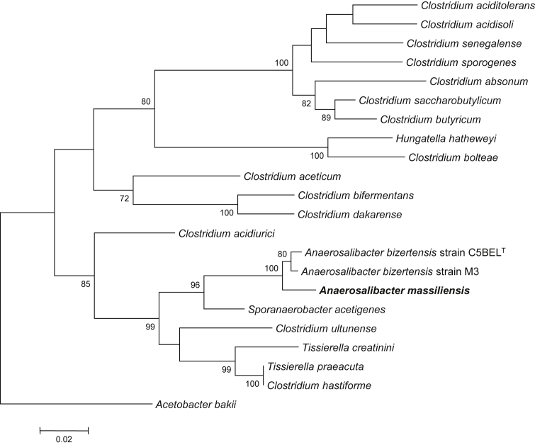
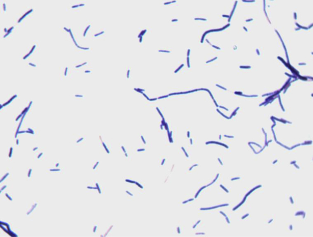
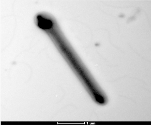
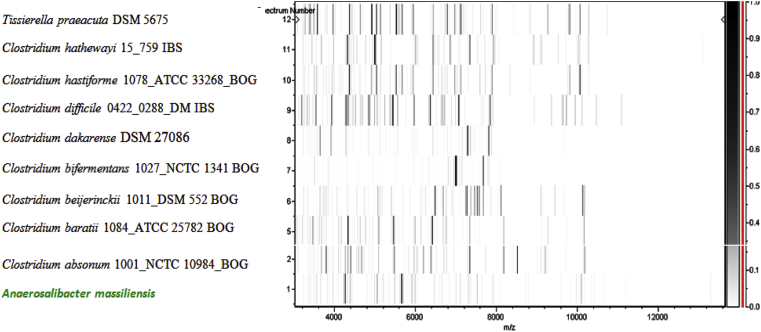
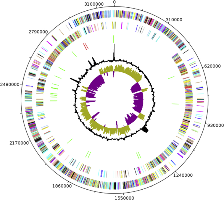
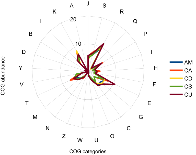
Anaerosalibacter massiliensis sp. nov. strain ND1(T) (= CSUR P762 = DSM 27308) is the type strain of A. massiliensis sp. nov., a new species within the genus Anaerosalibacter. This strain, the genome of which is described here, was isolated from the faecal flora of a 49-year-old healthy Brazilian man. Anaerosalibacter massiliensis is a Gram-positive, obligate anaerobic rod and member of the family Clostridiaceae. With the complete genome sequence and annotation, we describe here the features of this organism. The 3 197 911 bp long genome (one chromosome but no plasmid) contains 3271 protein-coding and 62 RNA genes, including six rRNA genes.

Keywords: Anaerosalibacter massiliensis; Clostridiaceae; culturomics; genome; taxonogenomics.


Figures

Fig. 1
Fig. 1
Phylogenetic tree highlighting position of Anaerosalibacter massiliensis sp. nov. strain ND1T relative to other type strains within Clostridiaceae. Strains and their corresponding GenBank accession numbers for 16S rRNA genes are (type = T): A. massiliensis strain ND1T, HG315673; A. bizertensis strain C5BELT, HQ534365; A. bizertensis strain M3, HG964477; S. acetigenes strain Lup33T, NR_025151 ; C. ultunense strain BST, GQ461825; T. creatinini strain BN11T, FR749955; C. hastiforme strain ATCC 33268T, X80841, ; T. preacuta strain ATCC 25539T, GQ461814; C. acidurici strain ATCC 7906T, M59084, ; C. aceticum strain ATCC 35044T, Y18183; C. bifermentans strain ATCC 638T, AB075769, ; C. dakarense strain FF1T, KC517358; C. saccharobutylicum strain ATCC BAA-117T, U16147; C. butyricum strain ATCC 19398T, AJ458420, ; C. absonum strain ATCC 27555T, X77842; C. senegalense strain JC122T, NR_125591 ; C. sporogenes strain ATCC 3584T, X68189, ; C. aciditolerans strain JW/YJL-B3T, DQ114945; C. acidisoli strain CK74T, AJ237756; Hungatella hatheweyi strain UB-B.2T, HE603919; C. bolteae strain ATCC BAA-613T, AJ508452, . 16S rRNA from A. massiliensis (1512 bp) was amplified and sequenced using fd1 (5′- AGAGTTTGATCCTGGCTCAG-3′) and rP2 (5′-ACGGCTACCTTGTTACGACTT-3′) primers; 16S rRNA sequences from all studied strains were aligned using CLUSTALW; total of 1182 nucleotide positions present in all studied sequences were used for phylogenetic inferences with maximum-likelihood method within MEGA6 software. Numbers at nodes are percentages of bootstrap values obtained by repeating analysis 500 times to generate majority consensus tree. Only bootstrap values greater than 70% are indicated. Acetobacterium bakii strain DSM 8239T, X96960, was used as outgroup. Scale bar = 2% nucleotide sequence divergence.
Fig. 2
Fig. 2
Gram staining of Anaerosalibacter massiliensis strain ND1T.
Fig. 3
Fig. 3
Transmission electron microscopy of Anaerosalibacter massiliensis strain ND1T using Morgani 268D (Philips, Amsterdam, The Netherlands) at operating voltage of 60 kV. Scale bar = 1 μm.
Fig. 4
Fig. 4
Reference mass spectrum from Anaerosalibacter massiliensis strain ND1T. This reference spectrum was generated by comparison of 12 individual colonies.
Fig. 5
Fig. 5
Gel view comparing Anaerosalibacter massiliensis strain ND1T to other members of family Clostridiaceae. Gel view displays raw spectra of loaded spectrum files arranged in pseudo-gel-like look. x-axis records m/z value. Left y-axis displays running spectrum number originating from subsequent spectra loading. Peak intensity is expressed as greyscale. The colour bar and right axis indicate the intensity each MALDI-TOF MS peak is displayed with and peak intensity in arbitrary units. Displayed species are detailed at left.
Fig. 6
Fig. 6
Graphical circular map of chromosome. From outside in, outer two circles show ORFs oriented in forward (coloured by COGs categories) and reverse (coloured by COGs categories) direction respectively. Third circle marks rRNA gene operon (red) and tRNA genes (green). Fourth circle shows G+C% content plot. Innermost circle shows GC skew, with purple indicating negative values and olive positive values.
Fig. 7
Fig. 7
Distribution of functional classes of predicted genes in genomes from Anaerosalibacter massiliensis (AM), Clostridium acidurici (CA), C. dakarense (CD), C. senegalense (CS) and C. ultunense (CU) chromosomes according to clusters of orthologous groups of proteins.

References

    1. Dubourg G., Lagier J.C., Armougom F., Robert C., Hamad I., Brouqui P. The gut microbiota of a patient with resistant tuberculosis is more comprehensively studied by culturomics than by metagenomics. Eur J Clin Microbiol Infect. 2013;32:637–645. - PubMed
    1. Lagier J.C., Armougom F., Million M., Hugon P., Pagnier I., Robert C. Microbial culturomics: paradigm shift in the human gut microbiome study. Clin Microbiol Infect. 2012;18:1185–1193. - PubMed
    1. Rezgui R., Maaroufi A., Fardeau M.L., Ben Ali Gam Z., Cayol J.L., Ben Hamed S. Anaerosalibacter bizertensis gen. nov., sp. nov., a halotolerant bacterium isolated from sludge. Int J Syst Evol Microbiol. 2014;62:2469–2474. - PubMed
    1. Ramasamy D., Mishra A.K., Lagier J.C., Padhmanabhan R., Rossi M., Sentausa E. A polyphasic strategy incorporating genomic data for the taxonomic description of new bacterial species. Int J Syst Evol Microbiol. 2014;64:384–391. - PubMed
    1. Lagier J.C., El Karkouri K., Nguyen T.T., Armougom F., Raoult D., Fournier P.E. Non-contiguous finished genome sequence and description of Anaerococcus senegalensis sp. nov. Stand Genomic Sci. 2012;6:116–125. - PMC - PubMed

LinkOut - more resources