

Beta-Catenin and Plakoglobin Expression during Zebrafish Tooth Development and Replacement
- PMID: 26938059
- PMCID: PMC4777446
- DOI: 10.1371/journal.pone.0148114
Beta-Catenin and Plakoglobin Expression during Zebrafish Tooth Development and Replacement
Abstract
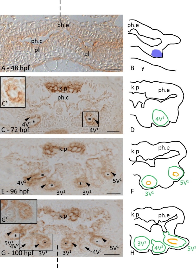
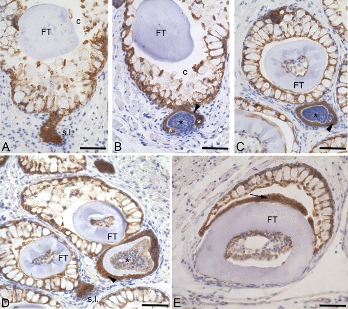
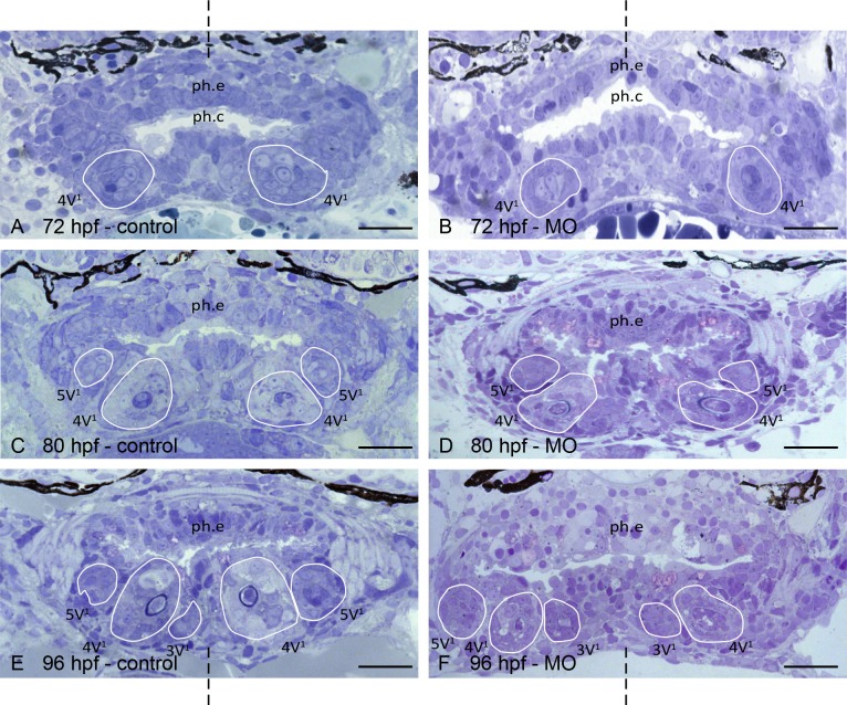
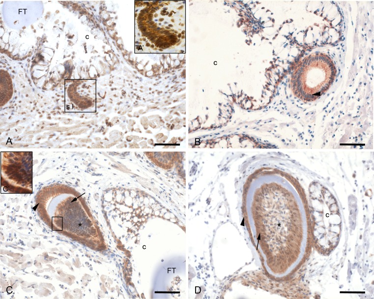
We analyzed the protein distribution of two cadherin-associated molecules, plakoglobin and β-catenin, during the different stages of tooth development and tooth replacement in zebrafish. Plakoglobin was detected at the plasma membrane already at the onset of tooth development in the epithelial cells of the tooth. This pattern remained unaltered during further tooth development. The mesenchymal cells only showed plakoglobin from cytodifferentiation onwards. Plakoglobin 1a morpholino-injected embryos showed normal tooth development with proper initiation and differentiation. Although plakoglobin is clearly present during normal odontogenesis, the loss of plakoglobin 1a does not influence tooth development. β-catenin was found at the cell borders of all cells of the successional lamina but also in the nuclei of surrounding mesenchymal cells. Only membranous, not nuclear, β-catenin, was found during morphogenesis stage. However, during cytodifferentiation stage, both nuclear and membrane-bound β-catenin was detected in the layers of the enamel organ as well as in the differentiating odontoblasts. Nuclear β-catenin is an indication of an activated Wnt pathway, therefore suggesting a possible role for Wnt signalling during zebrafish tooth development and replacement.
Conflict of interest statement
Figures
Similar articles
-
Enhanced Wnt/β-catenin signalling during tooth morphogenesis impedes cell differentiation and leads to alterations in the structure and mineralisation of the adult tooth.Biol Cell. 2012 Oct;104(10):603-17. doi: 10.1111/boc.201100075. Epub 2012 Aug 23. Biol Cell. 2012. PMID: 22671936
-
Plakoglobin expression and localization in zebrafish embryo development.Biochem Soc Trans. 2004 Nov;32(Pt 5):797-8. doi: 10.1042/BST0320797. Biochem Soc Trans. 2004. PMID: 15494018
-
N-cadherin is required for cytodifferentiation during zebrafish odontogenesis.J Dent Res. 2013 Apr;92(4):365-70. doi: 10.1177/0022034513477424. Epub 2013 Feb 8. J Dent Res. 2013. PMID: 23396519
-
The canonical way to make a heart: β-catenin and plakoglobin in heart development and remodeling.Exp Biol Med (Maywood). 2017 Dec;242(18):1735-1745. doi: 10.1177/1535370217732737. Epub 2017 Sep 18. Exp Biol Med (Maywood). 2017. PMID: 28920469 Free PMC article. Review.
-
Catenins, Wnt signaling and cancer.Bioessays. 2000 Nov;22(11):961-5. doi: 10.1002/1521-1878(200011)22:11<961::AID-BIES1>3.0.CO;2-T. Bioessays. 2000. PMID: 11056471 Review.
Cited by
-
Synergistic roles of Wnt modulators R-spondin2 and R-spondin3 in craniofacial morphogenesis and dental development.Sci Rep. 2021 Mar 12;11(1):5871. doi: 10.1038/s41598-021-85415-y. Sci Rep. 2021. PMID: 33712657 Free PMC article.
-
SHH signaling directed by two oral epithelium-specific enhancers controls tooth and oral development.Sci Rep. 2017 Oct 11;7(1):13004. doi: 10.1038/s41598-017-12532-y. Sci Rep. 2017. PMID: 29021530 Free PMC article.
-
Duplication and gene expression patterns of β-catenin in Nile tilapia.Fish Physiol Biochem. 2018 Apr;44(2):651-659. doi: 10.1007/s10695-017-0460-2. Epub 2017 Dec 30. Fish Physiol Biochem. 2018. PMID: 29290067
-
Mapping the Tooth Enamel Proteome and Amelogenin Phosphorylation Onto Mineralizing Porcine Tooth Crowns.Front Physiol. 2019 Jul 30;10:925. doi: 10.3389/fphys.2019.00925. eCollection 2019. Front Physiol. 2019. PMID: 31417410 Free PMC article.
-
Distinct tooth regeneration systems deploy a conserved battery of genes.Evodevo. 2021 Mar 25;12(1):4. doi: 10.1186/s13227-021-00172-3. Evodevo. 2021. PMID: 33766133 Free PMC article.
References
Publication types
MeSH terms
Substances
LinkOut - more resources
Full Text Sources
Other Literature Sources
Molecular Biology Databases
