. 2016 Mar 3;11(3):e0150635.
doi: 10.1371/journal.pone.0150635. eCollection 2016.

Foxn1 Transcription Factor Regulates Wound Healing of Skin through Promoting Epithelial-Mesenchymal Transition

Affiliations

Foxn1 Transcription Factor Regulates Wound Healing of Skin through Promoting Epithelial-Mesenchymal Transition

Barbara Gawronska-Kozak et al. PLoS One. .

Abstract

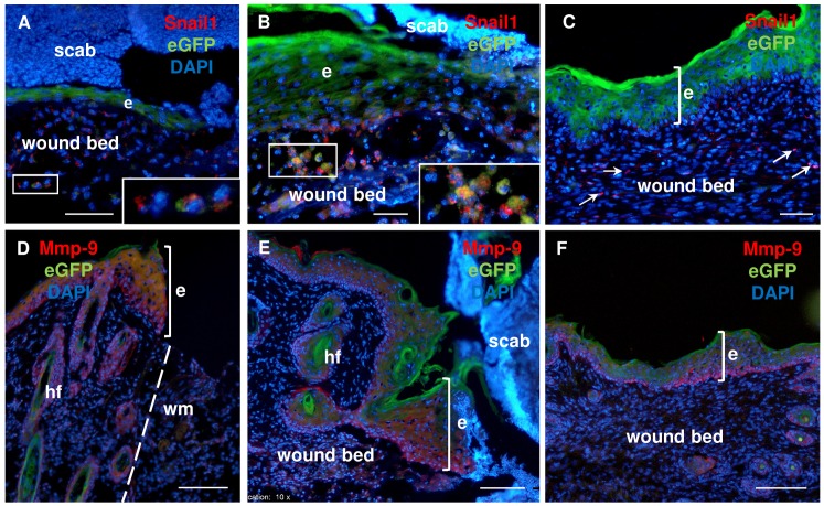
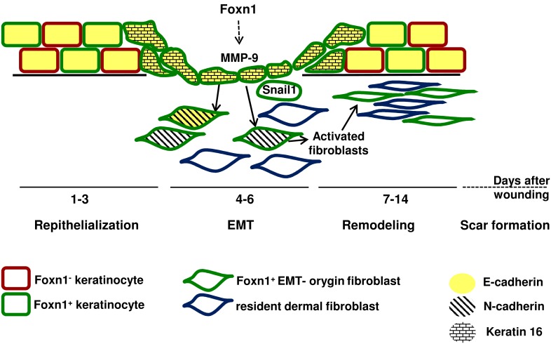
Transcription factors are key molecules that finely tune gene expression in response to injury. We focused on the role of a transcription factor, Foxn1, whose expression is limited to the skin and thymus epithelium. Our previous studies showed that Foxn1 inactivity in nude mice creates a pro-regenerative environment during skin wound healing. To explore the mechanistic role of Foxn1 in the skin wound healing process, we analyzed post-injured skin tissues from Foxn1::Egfp transgenic and C57BL/6 mice with Western Blotting, qRT-PCR, immunofluorescence and flow cytometric assays. Foxn1 expression in non-injured skin localized to the epidermis and hair follicles. Post-injured skin tissues showed an intense Foxn1-eGFP signal at the wound margin and in leading epithelial tongue, where it co-localized with keratin 16, a marker of activated keratinocytes. This data support the concept that suprabasal keratinocytes, expressing Foxn1, are key cells in the process of re-epithelialization. The occurrence of an epithelial-mesenchymal transition (EMT) was confirmed by high levels of Snail1 and Mmp-9 expression as well as through co-localization of vimentin/E-cadherin-positive cells in dermis tissue at four days post-wounding. Involvement of Foxn1 in the EMT process was verified by co-localization of Foxn1-eGFP cells with Snail1 in histological sections. Flow cytometric analysis showed the increase of double positive E-cadherin/N-cadherin cells within Foxn1-eGFP population of post-wounded skin cells isolates, which corroborated histological and gene expression analyses. Together, our findings indicate that Foxn1 acts as regulator of the skin wound healing process through engagement in re-epithelization and possible involvement in scar formation due to Foxn1 activity during the EMT process.


Conflict of interest statement

Competing Interests: The authors have declared that no competing interests exist.

Figures

Fig 1
Fig 1. Macroscopic and microscopic evaluation of the skin wound healing process in Foxn1::eGFP and B6 mice.
(A) Representative macroscopic views of skin wounds at days 1–7 and 14 after wounding. (B) The morphometrical analysis of the wound closure areas. (C) Representative histological sections of post-wounded skin area of B6 mice at day 3 (hematoxylin and eosin staining; (HE)); scale bar 500 μm. (D) The comparison of time-course of re-epithelialization process between Foxn1::Egfp and B6 mice. wm—wound margin; ne–newly formed epithelium delineated by dotted line. Values are the mean ± SEM; *p<0.05; ***p<0.001.
Fig 2
Fig 2
Spatial and temporal histological analysis of uninjured (A) and post-injured (B-G) skin from Foxn1::Egfp mice. Skin sections at post-wounded day 2 (B), day 3 (C, D), day 4 (E, F) and day 7 (G). Nuclei were counterstained with DAPI. hf–hair follicle, e–epidermis; g–granulation tissue; wm–wound margin; dotted line delineated basement membrane; arrow–indicates the direction of neo-epithelium migration. Scale bar: 50μm (A, F), 100 μm (B, C, E, G), 200 μm (D).
Fig 3
Fig 3. Foxn1-eGFP protein expression during the time course of skin wound healing in Foxn1::Egfp mice.
(A) Densitometric analysis of eGFP protein from n = 43 Foxn1::Egfp mice, with n = 4–6 single skin samples per time point. (B) Representative Western blot analysis of Foxn1-eGFP protein expression in skin tissues collected from Foxn1::Egfp mice Values are the mean ± SEM; ** p<0.01; *** p<0.001).
Fig 4
Fig 4
Immunofluorescent detection of E-cadherin (A–C) and keratin 16 (D–E) expression in uninjured (A) and post-injured at day 2 (B–D) and day 3 (E) skin tissues of Foxn1::Egfp mice. (A and B) E-cadherin, (A’ and B’) eGFP, (A” and B”) co-localization of E-cadherin and eGFP. hf–hair follicles; wm–wound margin; dotted line–basement membrane (a, a’ and a”); dashed lines–delineated epidermal basal cells (b, b’ and b”); arrow–neo-epithelium migration; e–epidermis; b–basal epidermal layer; s–suprabasal and squamous epidermal layers. Scale bar 50 μm (A-A”, B-B” and C); 100 μm (D-E).
Fig 5
Fig 5
Snail1 (A) and Mmp-9 (B) mRNA expression during the time course of skin wound healing in B6 mice. Expression of Snail1 (A) and Mmp-9 (B) mRNA was analyzed in single skin samples (n = 6–9 per time point) and were normalized by the levels of Tbp mRNA. Values are the mean ± SEM; * p<0.05; ** p<0.01; *** p<0.001).
Fig 6
Fig 6. Immunofluorescent detection of EMT traits during the skin wound healing process in B6 mice.
Confocal microscopy imaging (A and A’) of co-localization of E-cadherin and vimentin-positive cells at day 6 after injury; insets provide its higher magnification. Immunostaining for Snali1 and Col IV were detected at post-wounded days: 2 (B), 3 (C), 4 (D), and 5 (E); nuclei were counterstained with DAPI. hf–hair follicle; e–epidermis; d–dermis; wm–wound margin; bm/arrows at B, D and E–basement membrane; arrowheads–Snail1 positive cells between fragmented basement membrane. Scale bar 20 μm (A-A’), 50 μm (E) and 100 μm (B-D).
Fig 7
Fig 7. Fluorescent detection of Foxn1-eGFP and EMT markers during skin wound healing process in Foxn1::Egfp mice.
Foxn1-eGFP and Snail1 fluorescent detection at days: 2 (A), 4 (B), and 6 (C). Foxn1-eGFP and Mmp-9 fluorescent detection at days: 2 (D), 4 (E), and 6 (F). Nuclei were counterstained with DAPI. hf–hair follicle; e–epidermis; wm–wound margin; arrows–Snail1 positive cells in dermis; insets show higher magnification. Scale bar 50 μm (A-C), 100 μm (D-F).
Fig 8
Fig 8. Flow cytometry analysis of cells isolated from post-injured and uninjured skin area of Foxn1::eGFP mice at post-wounded day 5th.
(A) Representative FACS histograms show detection of Foxn1-eGFP-positive populations of cells from injured skin of Foxn1::Egfp (black) and B6 (grey) mice. (C) Foxn1-eGFP-positive population of cells in uninjured versus injured skin samples. (B, D) Analysis of E-cadherin and N-cadherin positive cells within Foxn1-eGFP population. Data represents mean ± SEM; n = 6.
Fig 9
Fig 9. Schematic illustrating the proposed role of Foxn1 in scar forming through involvement in EMT process.

References

    1. Slominski AT, Zmijewski MA, Skobowiat C, Zbytek B, Slominski RM, Steketee JD. Sensing the environment: regulation of local and global homeostasis by the skin's neuroendocrine system. Adv Anat Embryol Cell Biol. 2012;212:v, vii, 1–115. Epub 2012/08/17. - PMC - PubMed
    1. Gurtner GC, Werner S, Barrandon Y, Longaker MT. Wound repair and regeneration. Nature. 2008;453(7193):314–21. Epub 2008/05/16. doi: nature07039 [pii] 10.1038/nature07039 . - DOI - PubMed
    1. Schafer M, Werner S. Transcriptional control of wound repair. Annu Rev Cell Dev Biol. 2007;23:69–92. Epub 2007/05/04. 10.1146/annurev.cellbio.23.090506.123609 . - DOI - PubMed
    1. Bellavia G, Fasanaro P, Melchionna R, Capogrossi MC, Napolitano M. Transcriptional control of skin reepithelialization. J Dermatol Sci. 2014;73(1):3–9. Epub 2013/09/10. 10.1016/j.jdermsci.2013.08.007 S0923-1811(13)00282-X [pii]. . - DOI - PubMed
    1. Colwell AS, Longaker MT, Lorenz HP. Mammalian fetal organ regeneration. Adv Biochem Eng Biotechnol. 2005;93:83–100. . - PubMed

Publication types

MeSH terms