. 2016 Mar 3;11(3):e0150204.
doi: 10.1371/journal.pone.0150204. eCollection 2016.

Retinal Image Simulation of Subjective Refraction Techniques

Affiliations

Retinal Image Simulation of Subjective Refraction Techniques

Sara Perches et al. PLoS One. .

Abstract

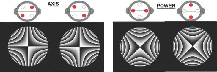
Refraction techniques make it possible to determine the most appropriate sphero-cylindrical lens prescription to achieve the best possible visual quality. Among these techniques, subjective refraction (i.e., patient's response-guided refraction) is the most commonly used approach. In this context, this paper's main goal is to present a simulation software that implements in a virtual manner various subjective-refraction techniques--including Jackson's Cross-Cylinder test (JCC)--relying all on the observation of computer-generated retinal images. This software has also been used to evaluate visual quality when the JCC test is performed in multifocal-contact-lens wearers. The results reveal this software's usefulness to simulate the retinal image quality that a particular visual compensation provides. Moreover, it can help to gain a deeper insight and to improve existing refraction techniques and it can be used for simulated training.


Conflict of interest statement

Competing Interests: The authors have declared that no competing interests exist.

Figures

Fig 1
Fig 1. Simulation software.
Main screen of the simulation software’s GUI.
Fig 2
Fig 2. Full process for retinal image computation.
Scheme of the procedure followed to generate the retinal image of a line of letters (0.20 logMAR). (a) Phase functions corresponding to the artificial eye (Weye), the phoropter’s lens (Wlens) and Jackson’s Cross Cylinder test lens (WJCC) (b) Sum of the three phase functions (WT (x,y)) and pupil function (P(x,y)), (c) Paraxial image affected by spectacle magnification, (d) PSF obtained in step (b) and (e) Retinal image, resulting from the convolution of the paraxial image and the PSF. Cross-cylinder power is ±0.50 D. Pupil radius is assumed to be 3 mm.
Fig 3
Fig 3. Simulated retinal images for a clock dial test.
Retinal images of a clock dial test computed for two subjects (P1 and P2) having very different aberration patterns. The images on the left (i.e., the first one of each row) correspond to the simulated retinal image when only spherical compensation has been virtually added. The remaining images correspond to the addition of the amount of cylinder power shown on top of each image, until equality was achieved. Pupil radius was 2 mm in all cases.
Fig 4
Fig 4. Jackson’s Cross Cylinder.
(A) A typical Jackson’s Cross Cylinder. Red lines indicate negative (minus) cylinder and black lines indicate positive (plus) cylinder. The handle points out the cross cylinder’s axis. (B) Section of the simulation software interface corresponding to Jackson’s Cross-Cylinder configuration. The pictures are used to determine the cylindrical correction axis (right) and power (left).
Fig 5
Fig 5. Interferometric images of Jackson’s Cross Cylinders.
Interferometric images shown in the simulation software representing the phase function for each position of the Jackson’s Cross-Cylinder (JCC). The image on the left is used to determine the cylindrical correction’s axis and the image on the right is used to determine the cylindrical power. Cross cylinder power is ±0.50 D. Pupil radius was 3 mm.
Fig 6
Fig 6. Simulated retinal images during JCC with a dot pattern.
Retinal images with a dot pattern as chart. On the left side, JCC test is performed while maintaining the CLC on the retina. On the right side, JCC test when CLC is placed elsewhere. The selected sphero-cylinder, once the compensation axis and cylindrical power had been determined, is indicated with a dashed line. Cross cylinder power is ±0.50 D. Pupil radius is 2 mm.
Fig 7
Fig 7. Simulated retinal images with the best compensation yielded by a JCC test where a dot pattern was used as chart.
Retinal images of a line of letters (0 logMAR) with the best sphero-cylindrical refraction that the JCC test yielded in two different scenarios: the CLC was maintained on the retina (A) and the CLC position was not closely controlled (B). Pupil radius was 2 mm.
Fig 8
Fig 8. Simulated retinal images during JCC test with a line of letters.
The selected sphero-cylinder, once the compensation axis and cylindrical power had been determined, is indicated with a dashed line. Cross cylinder power is ±0.50 D. Pupil radius was 2 mm. Visual acuity for the line of letters corresponds with 0.20 logMAR.
Fig 9
Fig 9. Simulated retinal images with the best compensation yielded by a JCC test where a line of letters was used as chart.
Retinal images of a line of letters (0 logMAR) with the best sphero-cylindrical refraction that the JCC test yielded in two different scenarios: the CLC was maintained on the retina (A) and the CLC position was not closely controlled (B). Pupil radius was 2 mm.
Fig 10
Fig 10. Multifocal concentric-ring contact lens designs.
CL1 is a centre-near (N) bifocal design contact lens with a distance zone (D) and intermediate-vision power (Int). CL2 is a centre-near bifocal design contact lens with no intermediate-vision zone.
Fig 11
Fig 11. Defocus curves.
Defocus curves for CL1 (black dots) and CL2 (red squares) multifocal contact lenses. The shaded area corresponds to the intermediate-vision zone. Pupil radius was 3 mm.
Fig 12
Fig 12. Through-focus simulated retinal images yielded by contact lens designs CL1 and CL2.
Retinal images obtained with CL1 and CL2, corresponding to a Snellen’s E with 0 logMAR located at different distances from the eye, going from more distant objects (left) to closer ones (right). The amount of defocus (in diopters) is indicated at the top of the figure. VS ratio is shown under each image. Pupil radius was 3 mm.
Fig 13
Fig 13. Simulated retinal images during a Jackson’s Cross Cylinder test with and without a multifocal contact lens.
Simulated retinal images produced during a JCC test for a subject wearing a multifocal contact lens (CL) (right column) or without CL (left column). Cross cylinder power was ±0.50 D and pupil radius was 3 mm.

References

    1. Benjamin WL. Borish’s Clinical Refraction Oxford: Butterworth-Heinemann; 2006.
    1. Bennett RB. Clinical Visual Optics. Oxford: Butterworth-Heinemann; 1998.
    1. Michaels DM. Visual Optics and Refraction: A Clinical Approach St Louis: The CV Mosby Co; 1985.
    1. Sims CN, Durham DG. The Jackson cross cylinder disproved. Trans Am Ophthalmol Soc. 1986;84:355–86. - PMC - PubMed
    1. Collins MJ, Shaw A, Menkens E, Davis B, Frankli R. The effect of pupil size on subjective refraction with irregular cornea. Invest Ophthalmol Vis Sci. 2002;43:E-Abstract 2058.

Publication types