. 2016 Mar 3;12(3):e1005476.
doi: 10.1371/journal.ppat.1005476. eCollection 2016 Mar.

Morphology and Molecular Composition of Purified Bovine Viral Diarrhea Virus Envelope

Affiliations

Morphology and Molecular Composition of Purified Bovine Viral Diarrhea Virus Envelope

Nathalie Callens et al. PLoS Pathog. .

Abstract

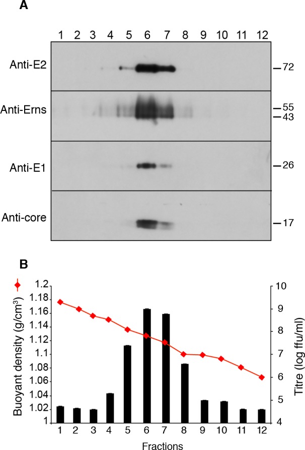
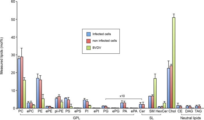
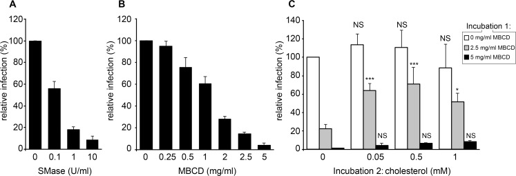
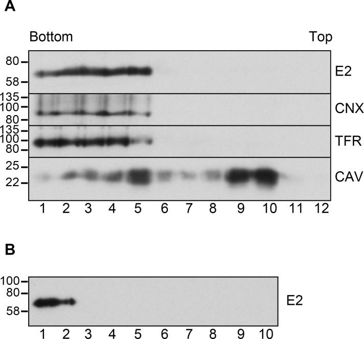
The family Flaviviridae includes viruses that have different virion structures and morphogenesis mechanisms. Most cellular and molecular studies have been so far performed with viruses of the Hepacivirus and Flavivirus genera. Here, we studied bovine viral diarrhea virus (BVDV), a member of the Pestivirus genus. We set up a method to purify BVDV virions and analyzed their morphology by electron microscopy and their protein and lipid composition by mass spectrometry. Cryo-electron microscopy showed near spherical viral particles displaying an electron-dense capsid surrounded by a phospholipid bilayer with no visible spikes. Most particles had a diameter of 50 nm and about 2% were larger with a diameter of up to 65 nm, suggesting some size flexibility during BVDV morphogenesis. Morphological and biochemical data suggested a low envelope glycoprotein content of BVDV particles, E1 and E2 being apparently less abundant than Erns. Lipid content of BVDV particles displayed a ~2.3 to 3.5-fold enrichment in cholesterol, sphingomyelin and hexosyl-ceramide, concomitant with a 1.5 to 5-fold reduction of all glycerophospholipid classes, as compared to lipid content of MDBK cells. Although BVDV buds in the endoplasmic reticulum, its lipid content differs from a typical endoplasmic reticulum membrane composition. This suggests that BVDV morphogenesis includes a mechanism of lipid sorting. Functional analyses confirmed the importance of cholesterol and sphingomyelin for BVDV entry. Surprisingly, despite a high cholesterol and sphingolipid content of BVDV envelope, E2 was not found in detergent-resistant membranes. Our results indicate that there are differences between the structure and molecular composition of viral particles of Flaviviruses, Pestiviruses and Hepaciviruses within the Flaviviridae family.


Conflict of interest statement

The authors have declared that no competing interests exist.

Figures

Fig 1
Fig 1. Flotation of BVDV particles in a sucrose density gradient.
BVDV was collected from culture medium of infected MDBK cells, concentrated by PEG precipitation, isolated in a 15–30% sucrose interface, adjusted to 40% sucrose, overlaid with a 5–35% sucrose density gradient and centrifuged. Fractions were collected from the bottom of the tube. (A) Immunoblot analysis of gradient fractions with indicated antibodies under reducing conditions for C, E1 and Erns, and non-reducing conditions for E2. (B) Infectious titers and buoyant density of gradient fractions.
Fig 2
Fig 2. Analysis of purified BVDV particles by electron microscopy.
Visualization of purified BVDV particles after negative staining (A, B), or by cryo-electron microscopy (C, D). Black arrows: enveloped, capsid-containing particles of ~50 nm, black arrowheads: larger enveloped, capsid-containing particles, white arrowheads: non-enveloped, capsid-like particles, white arrows: small empty vesicles, white asterisks: large vesicles. (E) Quantification of different objects indicated in C and D. (F) examples of images used for virion reconstruction. (G) Virion reconstructed by projecting 160 images of 50-nm particles. Bars, 200 nm (A) or 100 nm (B,D).
Fig 3
Fig 3. Immunocapture of BVDV particles.
Protein G-coated magnetic beads (asterisks) were incubated with anti-BVDV E2 mAbs (A), no antibody (B), or an irrelevant mAb (C) and purified BVDV fraction and observed by cryo-electron microscopy. Black arrows: enveloped, capsid-containing particles of ~50 nm, black arrowheads: larger enveloped, capsid-containing particles. Bar, 100 nm.
Fig 4
Fig 4. Envelope glycoprotein dimerization.
(A) Triton-X100 lysates of infected and non-infected MDBK cells analyzed by immunoblot with a mix of anti-E2 mAbs WB214 and WB166 under reduced or non-reduced conditions. (B) Purified BVDV analyzed by immunoblot with anti-E2 mAbs WB214 and WB166, anti-E1 mAb 8F2 or anti-Erns pAb under reduced (R) or non-reduced (NR) conditions. (C) Lysates of purified BVDV and infected MDBK cells containing similar amounts of E1E2 heterodimer, and a lysate of non-infected MDBK cells were analyzed by immunoblot with anti-E2 mAbs under non-reduced condition. The bands of E2 monomer, E2 homodimer and E1E2 heterodimer are indicated.
Fig 5
Fig 5. Protein content of purified BVDV fraction.
(A) A purified BVDV fraction and an equivalent fraction obtained from non-infected cells (control) were analyzed by SDS-PAGE under reducing conditions and colloidal Coomassie blue staining. Asterisk, Erns, double asterisk, core. (B) Immunoblot analysis under reducing conditions and Ponceau red staining of purified BVDV fraction. (C) Distribution of CD9, annexin A2, ALIX and BVDV E2 in sucrose density gradient of BVDV (top) and control (bottom) fractions. Bands were quantified by densitometry and plotted over fraction density. (D) Immunoblot analysis of annexin A2 and BVDV core in flow through (1), PBS washes (2 and 3) and 0.4M NaCl elution (4 and 5) fractions of cellulose-sulfate column. 1:50 of each fraction was loaded on the gel. (E) Immunoblot analysis of Annexin A2, ALIX, CD9 and core in BVDV fractions before and after cellulose sulfate chromatography. 1:20 of input and eluted fraction were loaded on the gel.
Fig 6
Fig 6. Lipid class composition of BVDV and MDBK cells.
The content of individual lipid classes of infected MDBK cells, non-infected MDBK cells and purified BVDV were standardized to mole percentage of all membrane lipids within the sample. Amounts of lipids measured in control preparation from uninfected cells were subtracted from BVDV lipid values. Glycerophospholipids (GPL): phosphatidic acid (PA), phosphatidylcholine/-ethanolamine/-glycerol/-inositol/-serine (PC/PE/PG/PI/PS), ether linked GPL (ePC/ePE/ePG/ePI/ePS/ePA), and ethanolamine plasmalogen (pl-PE). Sphingolipids (SL): ceramide (Cer), sphingomyelin (SM) and hexosylceramide (HexCer). Sterols: cholesterol (Chol). Storage neutral lipids: cholesteryl esters (CE), diacylglycerol (DAG) and triacylglycerol (TAG). Error bars correspond to SDs (n = 3).
Fig 7
Fig 7. Phosphatidylcholine species of BVDV and MDBK cells.
(A) PC saturation as a number of double bonds in both fatty acyl chains. (B) PC chain length as number of carbon atoms in both fatty acyl chains. (C) Molecular PC species. Amounts of each lipid were standardized to the total identified PC species. Error bars correspond to SDs (n = 3).
Fig 8
Fig 8. Lipid compositions of viruses.
(A) Major lipid categories and (B) glycerophospholipid class compositions of influenza virus (IFV, ref #30), vesicular stomatitis virus (VSV), Semliki forest virus (SFV) (both taken from ref #29), human immunodeficiency virus (HIV, ref #28), hepatitis C virus (HCV, ref #6) and BVDV (this study). GPL: glycerophospholipid, SL: sphingolipid, chol: cholesterol, PI: phosphatidylinositol, PS phosphatidylserine, PE: phosphatidylethanolamine, PC: phosphatidylcholine, ePE: ether-linked PE, ePC: ether-linked PC. Cholesteryl esters of HCV have been omitted to facilitate the comparison with other viruses.
Fig 9
Fig 9. Sphingomyelin and cholesterol in BVDV entry.
(A) Unpurified BVDV was incubated for 1h at 37°C in the presence of indicated concentrations of sphingomyelinase (SMase), diluted 1,000 times and used to infect MDBK cells. (B) Partially purified BVDV was incubated at 37°C for 1 h with indicated concentrations of methyl-β-cyclodextrin (MBCD), diluted 10,000 times and used to infect MDBK cells. (C) Partially purified BVDV was incubated at 37°C for 1 h with indicated concentrations of MBCD (incubation 1), diluted 100 times, incubated for 1h at 37°C with MBCD:cholesterol complex at final cholesterol concentrations indicated (incubation 2), diluted 100 times and used to infect MDBK cells. The number of infected cells was measured using an immunofluorescence assay at 15 hpi and standardized to the number of cells infected with untreated virus. Error bars correspond to SDs (n = 3, * P<0.05, *** P<0.001, NS: P>0.05; cholesterol-treated vs corresponding control, 2-way ANOVA).
Fig 10
Fig 10. E2 is excluded from detergent-resistant membranes.
DRMs were isolated by cold extraction of BVDV-infected MDBK cells (A) or partially purified BVDV (B) with Triton-X100 and flotation as explained in the Materials and Methods section. The distribution of BVDV E2, calnexin(CNX), transferrin receptor (TFR) and caveolin (CAV) was determined by immunoblot under reducing (CNX, TFR) or non-reducing (E2, CAV) conditions.

Similar articles

Cited by

References

    1. Lindenbach BD, Thiel H-J, Rice CM. Flaviviridae: the viruses and their replication. Knipe DM, Howley PM, editors. Fields Virology. 5 ed. Philadelphia; 2007;: 1101–1152.
    1. Rey FA, Heinz FX, Mandl C, Kunz C, Harrison SC. The envelope glycoprotein from tick-borne encephalitis virus at 2 A resolution. Nature. 1995;375: 291–298. 10.1038/375291a0 - DOI - PubMed
    1. Kuhn RJ, Zhang W, Rossmann MG, Pletnev SV, Corver J, Lenches E, et al. Structure of dengue virus: implications for flavivirus organization, maturation, and fusion. Cell. 2002;108: 717–725. - PMC - PubMed
    1. Bartenschlager R, Penin F, Lohmann V, André P. Assembly of infectious hepatitis C virus particles. Trends Microbiol. 2011;19: 95–103. 10.1016/j.tim.2010.11.005 - DOI - PubMed
    1. Lindenbach BD, Rice CM. The ins and outs of hepatitis C virus entry and assembly. Nat Rev Micro. 2013;11: 688–700. 10.1038/nrmicro3098 - DOI - PMC - PubMed

Publication types

MeSH terms

LinkOut - more resources