

Effect of intravitreal dexamethasone implant on retinal and choroidal thickness in refractory diabetic macular oedema after multiple anti-VEGF injections
- PMID: 26939558
- PMCID: PMC4869136
- DOI: 10.1038/eye.2016.23
Effect of intravitreal dexamethasone implant on retinal and choroidal thickness in refractory diabetic macular oedema after multiple anti-VEGF injections
Abstract
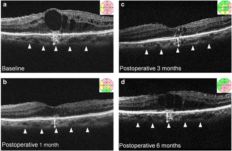
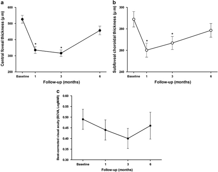
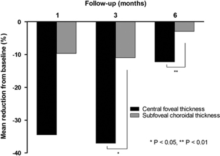
AimsTo investigate the effect of intravitreal dexamethasone implant (IVD) on central foveal thickness (CFT), choroidal thickness (CT) and its correlation with visual acuity in eyes with refractory diabetic macular oedema (DMO).MethodsThis was a retrospective interventional case-series. Thirty-five eyes of 35 patients were treated with a single injection of IVD because of refractory DMO with CFT over 300 μm, and persistent intraretinal and subretinal fluid despite of multiple intravitreal bevacizumab injections. Patients were followed-up for 6 months for the evaluation of CFT and subfoveal CT by spectral-domain optical coherence tomography.ResultsAll eyes (mean age: 59.4±12.35 years; 18 males, 17 females) had been previously treated with multiple bevacizumab injections and showed persistent DMO (mean number of injections 4.08±2.98) The preoperative logMAR BCVA was 0.49±0.24, which gradually improved to 0.46±0.32 at 6 months (P=0.652) and 26% gained two or more lines of Snellen visual acuity. At baseline, the mean CFT was 526.29±123.48 μm, which significantly improved to 316.15±100.09 μm at 3 months (P<0.001). However, CFT deteriorated to 457.07±136.53 μm at 6 months (P=0.051). Similarly, the mean preoperative subfoveal CT was 288.91±36.47 μm and it decreased to 266.85±30.93 μm at 3 months (P<0.01), but increased to 278.63±32.55 μm at 6 months (P=0.137). The reduction of CFT from baseline showed significant correlation with that of subfoveal CT at 3 months (P=0.041) and at 6 months (P=0.008).ConclusionsIn DMO refractory to multiple bevacizumab injections, IVD significantly reduced CFT and subfoveal CT, with BCVA improvement in one-fourth of the patients. The reduction of CFT showed significant correlation with reduction of subfoveal CT.
Figures
References
-
- Antonetti DA, Klein R, Gardner TW. Diabetic retinopathy. N Engl J Med 2012; 366(13): 1227–1239. - PubMed
-
- Adamis AP, Miller JW, Bernal MT, D'Amico DJ, Folkman J, Yeo TK et al. Increased vascular endothelial growth factor levels in the vitreous of eyes with proliferative diabetic retinopathy. Am J Ophthalmol 1994; 118(4): 445–450. - PubMed
-
- Arimura N, Otsuka H, Yamakiri K, Sonoda Y, Nakao S, Noda Y et al. Vitreous mediators after intravitreal bevacizumab or triamcinolone acetonide in eyes with proliferative diabetic retinopathy. Ophthalmology 2009; 116(5): 921–926. - PubMed
-
- Funatsu H, Noma H, Mimura T, Eguchi S. Vitreous inflammatory factors and macular oedema. Br J Ophthalmol 2012; 96(2): 302–304. - PubMed
-
- Funatsu H, Noma H, Mimura T, Eguchi S, Hori S. Association of vitreous inflammatory factors with diabetic macular edema. Ophthalmology 2009; 116(1): 73–79. - PubMed
Publication types
MeSH terms
Substances
LinkOut - more resources
Full Text Sources
Other Literature Sources
Medical
