Review
. 2016;11(3):441-53.
doi: 10.2217/fmb.15.151. Epub 2016 Mar 4.

Histones as mediators of host defense, inflammation and thrombosis

Affiliations
Review

Histones as mediators of host defense, inflammation and thrombosis

Marloes Hoeksema et al. Future Microbiol. 2016.

Abstract

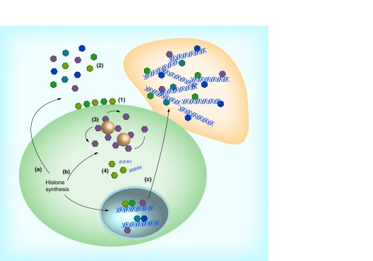
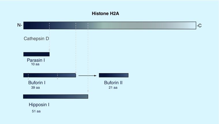
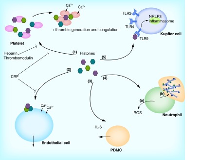
Histones are known for their ability to bind to and regulate expression of DNA. However, histones are also present in cytoplasm and extracellular fluids where they serve host defense functions and promote inflammatory responses. Histones are a major component of neutrophil extracellular traps that contribute to bacterial killing but also to inflammatory injury. Histones can act as antimicrobial peptides and directly kill bacteria, fungi, parasites and viruses, in vitro and in a variety of animal hosts. In addition, histones can trigger inflammatory responses in some cases acting through Toll-like receptors or inflammasome pathways. Extracellular histones mediate organ injury (lung, liver), sepsis physiology, thrombocytopenia and thrombin generation and some proteins can bind histones and reduce these potentially harmful effects.

Keywords: antimicrobial peptides; histones; innate immunity; neutrophils; platelets.


Conflict of interest statement

Financial & competing interests disclosure

The authors have no relevant affiliations or financial involvement with any organization or entity with a financial interest in or financial conflict with the subject matter or materials discussed in the manuscript. This includes employment, consultancies, honoraria, stock ownership or options, expert testimony, grants or patents received or pending, or royalties.

No writing assistance was utilized in the production of this manuscript.

Figures

<b>Figure 1.</b>
Figure 1.. Antimicrobial action of histones.
After synthesis in the cytoplasm, histones are transported to the nucleus where they regulate DNA condensation and gene transcription. Alternatively, histones can have extranuclear functions, either in the cytoplasm (b) or in the extracellular space (a). Histones transported from the cytoplasm either remain membrane-bound (1) or are released into the extracellular space (2), where they exert broad-spectrum antimicrobial activity against bacteria, viruses, parasites and fungi. Within the cytoplasm, histone H1 is bound to lipid droplets and is released upon stimulation with endotoxin or lipotechoic acid (3). In addition, histone H2B functions as a sensor for viral dsDNA (4). Nuclear histones end up in neutrophil extracellular traps (c), where they play an important part in neutrophil extracellular trap-mediated bacterial killing.
<b>Figure 2.</b>
Figure 2.. Cleavage products of histone H2A.
Cleavage products of histone H2A have been found in a number of species. In Parasilus asotus, an N-terminal 19aa fragment known as Parasin I is cleaved from full-length H2A by cathepsin D upon epidermal injury. Buforin I has been identified as a 39aa cleavage product and can be isolated from the stomach of Bufo bufo gargarizans. A smaller, 21aa peptide can be derived from Buforin I. A larger N-terminal fragment, hipposin I, was found in Hippoglossus hippoglossus L.
<b>Figure 3.</b>
Figure 3.. Mechanisms of histone-induced cellular damage.
When released from cells, histones can damage host cells and modulate inflammatory responses. Histones can bind to platelets (1), which results in calcium influx, platelet activation and aggregation. In addition, histones induce coagulation and the generation of thrombin. Histone-mediated injury can be reduced by administration of heparin and thrombomodulin, as well as CRP, which binds directly to histones. Direct cellular toxic effects can be observed in endothelial cells (2). Histones bind to the endothelial cell membrane, resulting in cell permeabilization, calcium influx and cell death. This can be inhibited by CRP. In addition, histones can activate phagocytic cells (3–5). PBMCs can be triggered to secrete IL-6 (3). When neutrophils are stimulated with histones, neutrophils release ROS as part of the neutrophil respiratory burst response (4a). Moreover, histones trigger NET formation in neutrophils (4b). Last, histones mediate activation of NRLP3 inflammasomes in Kupffer cells (5), which results in sterile liver injury. This inflammasome activation is thought to be activated through binding of histones to TLR9. In addition, histones can also activate TLR2 and TLR4. ROS: Reactive oxygen species.

References

    1. Luger K, Mader AW, Richmond RK, Sargent DF, Richmond TJ. Crystal structure of the nucleosome core particle at 2.8 A resolution. Nature. 1997;389(6648):251–260. - PubMed
    1. Allan J, Hartman PG, Crane-Robinson C, Aviles FX. The structure of histone H1 and its location in chromatin. Nature. 1980;288(5792):675–679. - PubMed
    1. Delange RJ, Smith EL. Histones: structure and function. Annu. Rev. Biochem. 1971;40:279–314. - PubMed
    1. Arents G, Burlingame RW, Wang BC, Love WE, Moudrianakis EN. The nucleosomal core histone octamer at 3.1 A resolution: a tripartite protein assembly and a left-handed superhelix. Proc. Natl Acad. Sci. USA. 1991;88(22):10148–10152. - PMC - PubMed
    1. Verdone L, Caserta M, Di Mauro E. Role of histone acetylation in the control of gene expression. Biochem. Cell Biol. 2005;83(3):344–353. - PubMed