Review
. 2016 May 1;310(9):G645-58.
doi: 10.1152/ajpgi.00015.2016. Epub 2016 Mar 3.

Glucosensing in the gastrointestinal tract: Impact on glucose metabolism

Affiliations
Review

Glucosensing in the gastrointestinal tract: Impact on glucose metabolism

Audren Fournel et al. Am J Physiol Gastrointest Liver Physiol. .

Abstract

The gastrointestinal tract is an important interface of exchange between ingested food and the body. Glucose is one of the major dietary sources of energy. All along the gastrointestinal tube, e.g., the oral cavity, small intestine, pancreas, and portal vein, specialized cells referred to as glucosensors detect variations in glucose levels. In response to this glucose detection, these cells send hormonal and neuronal messages to tissues involved in glucose metabolism to regulate glycemia. The gastrointestinal tract continuously communicates with the brain, especially with the hypothalamus, via the gut-brain axis. It is now well established that the cross talk between the gut and the brain is of crucial importance in the control of glucose homeostasis. In addition to receiving glucosensing information from the gut, the hypothalamus may also directly sense glucose. Indeed, the hypothalamus contains glucose-sensitive cells that regulate glucose homeostasis by sending signals to peripheral tissues via the autonomous nervous system. This review summarizes the mechanisms by which glucosensors along the gastrointestinal tract detect glucose, as well as the results of such detection in the whole body, including the hypothalamus. We also highlight how disturbances in the glucosensing process may lead to metabolic disorders such as type 2 diabetes. A better understanding of the pathways regulating glucose homeostasis will further facilitate the development of novel therapeutic strategies for the treatment of metabolic diseases.

Keywords: diabetes; glucose homeostasis; glucosensing.


Figures

Fig. 1.
Fig. 1.
Molecular mechanisms involved in glucosensing in the oral cavity. In the taste buds, activated TAS1R2/TAS1R3 heterodimers interact with G proteins comprising α-gustducin, leading to phospholipase C-β2 (PLC-β2) activation. In turn, activated PLC-β2 cleaves phosphatidylinositol 4,5-bisphosphate (PIP2) into inositol 1,4,5-trisphosphate (IP3) and diacylglycerol (DAG). IP3 stimulates Ca2+ release from the endoplasmic reticulum via type III IP3 receptor activation (IP3R). The elevated intracellular Ca2+ activates the transient receptor potential cation channel subfamily M member 5 (TRPM5) and induces membrane depolarization and ATP release, which stimulates efferent nerve fibers. GLUT-2/5, glucose transporter-2/5; SGLT-1, sodium-glucose cotransporter-1.
Fig. 2.
Fig. 2.
Molecular mechanisms involved in glucosensing in the gastrointestinal tract. A: in enterocytes, transport of glucose occurs by SGLT-1, following the Na+ electrochemical potential gradient across the apical membrane until its intracellular concentration is sufficiently elevated. Glucose is transported outside of enterocytes by facilitated diffusion through GLUT-2, localized on the basolateral membrane of the cell. Saturation of SGLT-1, depending on the luminal glucose concentration, is associated with an increase of GLUT-2 translocation to the apical membrane. B: in brush cells, the glucose trafficking signal transduction pathway involves GLUT-2 transporters. This mechanism is coupled with TAS1R2/TAS1R3 sweet taste receptor activation that was described for taste buds. C: enteroendocrine cells (EEC) receive glucose stimulation via sweet taste receptor activation, as described below, but GLUT-2 also trigger the entry of glucose into the glycolytic pathway to generate ATP. This increase in ATP leads to closure of ATP sensitive potassium channels (KATP) channels, membrane depolarization, opening of Ca2+ channels, and release of hormones like glucose-dependent insulinotropic polypeptide (GIP) and glucagon-like peptide-1 (GLP-1); note that membrane depolarization can be induced by glucose itself. D: enteric neurons are glucosensitive: extracellular glucose removal leads to hyperpolarization and decreases in their membrane input resistance. The excitation of these neurons is mediated by inhibition of KATP channels or by SGLT-3. They can also be activated by gastrointestinal (GI) hormones secreted by EECs in response to glucose detection. Enteric neurons can activate neighboring neurons or transmit glycemic information to the brain via nervous afferents.
Fig. 3.
Fig. 3.
Molecular mechanisms involved in glucosensing in the pancreas. In pancreatic β-cells, glucose is transported by GLUT-2, phosphorylated to glucose 6-phosphate by hexokinase IV, and subjected to the glycolysis pathway. This leads to an increase in ATP, followed by KATP channel closure and membrane depolarization. This depolarization, in turn, activates voltage-dependent Ca2+ channels, leading to Ca2+ accumulation in the cytoplasm and insulin secretion.
Fig. 4.
Fig. 4.
Molecular mechanisms involved in glucosensing in the portal vein. In hepatoportal glucosensor cells, portal glucose concentrations are inversely correlated with the firing rate of hepatic afferent nerves. Little is known about the molecular mechanisms involved in hepatoportal glucosensing, but several actors are required, including GLUT-2, GLUT-4, AMP-activated protein kinase (AMPK), and GLP-1 receptor.
Fig. 5.
Fig. 5.
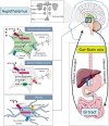
The gut-brain axis and the key role of hypothalamic glucosensing. Top, schematic representation of a coronal section showing different hypothalamic nuclei, including the arcuate nucleus (ARC), ventromedial nuclei (VMN), dorsomedial nuclei (DMN), the lateral hypothalamus nucleus (LH), and paraventricular nuclei (PVN). The remainder of the figure highlights molecular mechanisms in different hypothalamic cell types involved in glucosensing, which contribute to glucose homeostasis via the gut-brain axis. In glucose-excited neurons, glucose enters via GLUT-2, is converted to glucose 6-phosphate by glucokinase, and is then used in the glycolytic pathway to generate ATP. The resultant increase in intracellular ATP levels inactivates KATP channels, leading to membrane depolarization and opening of Ca2+ channels. The massive influx of Ca2+ causes release of neurotransmitters. Conversely, glucose-inhibited neurons are sensitive to decreases in the glucose concentration. Here, less glucose enters the neurons through GLUT-2 and is metabolized by the glycolytic pathway, thus producing less cellular ATP. The decrease in the ATP-to-ADP ratio is detected by AMPK, which activates production of gaseous messenger nitric oxide (NO) by neuronal NO synthase (nNOS). Production of NO leads to the inhibition of specific chloride channels, including the cystic fibrosis transmembrane regulator (CFTR), which is responsible for membrane depolarization. In glial cells, glucose enters into intracellular compartments via GLUT-2 and increases intracellular Ca2+ waves, a mechanism dependent on ATP release by connexin 43.

References

    1. Adachi A, Shimizu N, Oomura Y, Kobashi M. Convergence of hepatoportal glucose-sensitive afferent signals to glucose-sensitive units within the nucleus of the solitary tract. Neurosci Lett 46: 215–218, 1984. - PubMed
    1. Ahren B. Autonomic regulation of islet hormone secretion–implications for health and disease. Diabetologia 43: 393–410, 2000. - PubMed
    1. Ahren B. Glucagon–early breakthroughs and recent discoveries. Peptides 67: 74–81, 2015. - PubMed
    1. Amato A, Cinci L, Rotondo A, Serio R, Faussone-Pellegrini MS, Vannucchi MG, Mule F. Peripheral motor action of glucagon-like peptide-1 through enteric neuronal receptors. Neurogastroenterol Motil 22: 664–e203, 2010. - PubMed
    1. Balkan B, Li X. Portal GLP-1 administration in rats augments the insulin response to glucose via neuronal mechanisms. Am J Physiol Regul Integr Comp Physiol 279: R1449–R1454, 2000. - PubMed

Publication types

LinkOut - more resources