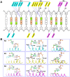
Structures of an all-α protein running along the DNA major groove
- PMID: 26939889
- PMCID: PMC4856987
- DOI: 10.1093/nar/gkw133
Structures of an all-α protein running along the DNA major groove
Abstract
Despite over 3300 protein-DNA complex structures have been reported in the past decades, there remain some unknown recognition patterns between protein and target DNA. The silkgland-specific transcription factor FMBP-1 from the silkworm Bombyx mori contains a unique DNA-binding domain of four tandem STPRs, namely the score and three amino acid peptide repeats. Here we report three structures of this STPR domain (termed BmSTPR) in complex with DNA of various lengths. In the presence of target DNA, BmSTPR adopts a zig-zag structure of three or four tandem α-helices that run along the major groove of DNA. Structural analyses combined with binding assays indicate BmSTPR prefers the AT-rich sequences, with each α-helix covering a DNA sequence of 4 bp. The successive AT-rich DNAs adopt a wider major groove, which is in complementary in shape and size to the tandem α-helices of BmSTPR. Substitutions of DNA sequences and affinity comparison further prove that BmSTPR recognizes the major groove mainly via shape readout. Multiple-sequence alignment suggests this unique DNA-binding pattern should be highly conserved for the STPR domain containing proteins which are widespread in animals. Together, our findings provide structural insights into the specific interactions between a novel DNA-binding protein and a unique deformed B-DNA.
© The Author(s) 2016. Published by Oxford University Press on behalf of Nucleic Acids Research.
Figures


Similar articles
-
DNA-binding property of the novel DNA-binding domain STPR in FMBP-1 of the silkworm Bombyx mori.J Biochem. 2009 Jul;146(1):103-11. doi: 10.1093/jb/mvp053. Epub 2009 Mar 20. J Biochem. 2009. PMID: 19304790
-
Structural approach to a novel tandem repeat DNA-binding domain, STPR, by CD and NMR.Biochemistry. 2007 Feb 20;46(7):1703-13. doi: 10.1021/bi061780c. Epub 2007 Jan 24. Biochemistry. 2007. PMID: 17249695
-
Structural properties of the DNA-bound form of a novel tandem repeat DNA-binding domain, STPR.Proteins. 2008 Jul;72(1):414-26. doi: 10.1002/prot.21939. Proteins. 2008. PMID: 18214959
-
Minor groove-binding architectural proteins: structure, function, and DNA recognition.Annu Rev Biophys Biomol Struct. 1998;27:105-31. doi: 10.1146/annurev.biophys.27.1.105. Annu Rev Biophys Biomol Struct. 1998. PMID: 9646864 Free PMC article. Review.
-
X-ray crystallographic studies of eukaryotic transcription initiation factors.Philos Trans R Soc Lond B Biol Sci. 1996 Apr 29;351(1339):483-9. doi: 10.1098/rstb.1996.0046. Philos Trans R Soc Lond B Biol Sci. 1996. PMID: 8735270 Review.
Cited by
-
Discovery of numerous novel Helitron-like elements in eukaryote genomes using HELIANO.Nucleic Acids Res. 2024 Sep 23;52(17):e79. doi: 10.1093/nar/gkae679. Nucleic Acids Res. 2024. PMID: 39119924 Free PMC article.
-
The large bat Helitron DNA transposase forms a compact monomeric assembly that buries and protects its covalently bound 5'-transposon end.Mol Cell. 2021 Oct 21;81(20):4271-4286.e4. doi: 10.1016/j.molcel.2021.07.028. Epub 2021 Aug 16. Mol Cell. 2021. PMID: 34403695 Free PMC article.
-
Protocols for protein-DNA binding analysis of a zinc finger transcription factor bound to its cognate promoter.STAR Protoc. 2022 Jul 31;3(3):101598. doi: 10.1016/j.xpro.2022.101598. eCollection 2022 Sep 16. STAR Protoc. 2022. PMID: 35928006 Free PMC article.
-
So close yet so far apart: distinct flanking sequence recognition by DNMT3A and DNMT3B.Commun Biol. 2025 Aug 13;8(1):1217. doi: 10.1038/s42003-025-08606-7. Commun Biol. 2025. PMID: 40804302 Free PMC article.
References
-
- Anderson J.E., Ptashne M., Harrison S.C. Structure of the repressor-operator complex of bacteriophage 434. Nature. 1987;326:846–852. - PubMed
-
- Matthews B.W. Protein-DNA interaction. No code for recognition. Nature. 1988;335:294–295. - PubMed
-
- Pabo C.O., Aggarwal A.K., Jordan S.R., Beamer L.J., Obeysekare U.R., Harrison S.C. Conserved residues make similar contacts in two repressor-operator complexes. Science. 1990;247:1210–1213. - PubMed
-
- Pavletich N.P., Pabo C.O. Crystal structure of a five-finger GLI-DNA complex: new perspectives on zinc fingers. Science. 1993;261:1701–1707. - PubMed
Publication types
MeSH terms
Substances
LinkOut - more resources
Full Text Sources
Other Literature Sources
Miscellaneous

