The NLRP3 and NLRP1 inflammasomes are activated in Alzheimer's disease
- PMID: 26939933
- PMCID: PMC4778358
- DOI: 10.1186/s13024-016-0088-1
The NLRP3 and NLRP1 inflammasomes are activated in Alzheimer's disease
Abstract
Background: Interleukin-1 beta (IL-1β) and its key regulator, the inflammasome, are suspected to play a role in the neuroinflammation observed in Alzheimer's disease (AD); no conclusive data are nevertheless available in AD patients.
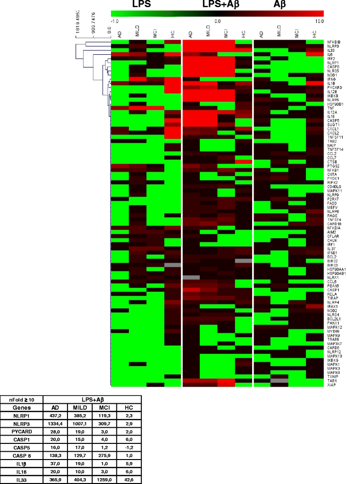
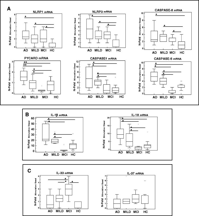
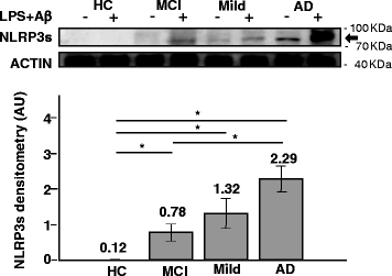
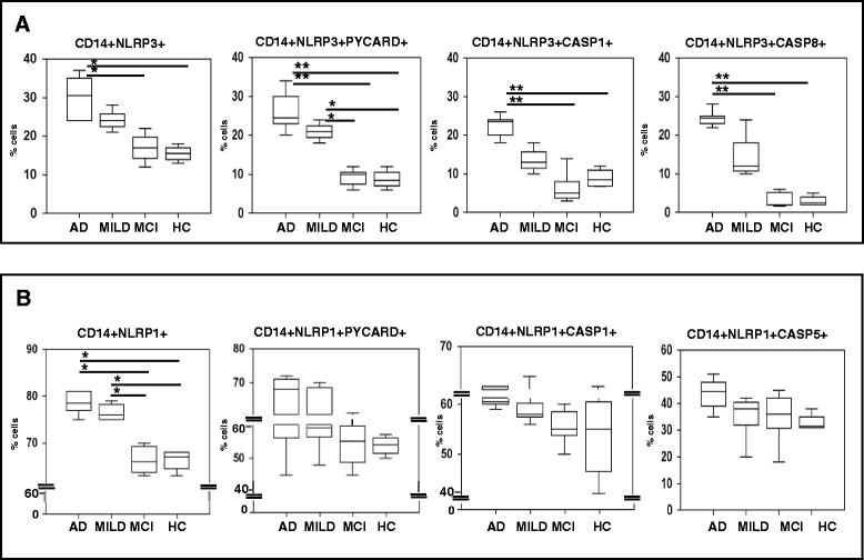
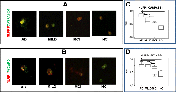
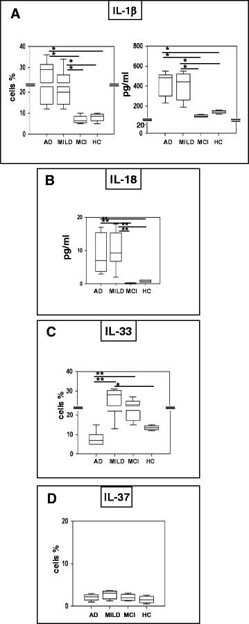
Results: mRNA for inflammasome components (NLRP1, NLRP3, PYCARD, caspase 1, 5 and 8) and downstream effectors (IL-1β, IL-18) was up-regulated in severe and MILD AD. Monocytes co-expressing NLRP3 with caspase 1 or caspase 8 were significantly increased in severe AD alone, whereas those co-expressing NLRP1 and NLRP3 with PYCARD were augmented in both severe and MILD AD. Activation of the NLRP1 and NLRP3 inflammasomes in AD was confirmed by confocal microscopy proteins co-localization and by the significantly higher amounts of the pro-inflammatory cytokines IL-1β and IL-18 being produced by monocytes. In MCI, the expression of NLRP3, but not the one of PYCARD or caspase 1 was increased, indicating that functional inflammasomes are not assembled in these individuals: this was confirmed by lack of co-localization and of proinflammatory cytokines production.
Conclusions: The activation of at least two different inflammasome complexes explains AD-associated neuroinflammation. Strategies targeting inflammasome activation could be useful in the therapy of AD.
Figures
Similar articles
-
Cytokine Secretion and Pyroptosis of Thyroid Follicular Cells Mediated by Enhanced NLRP3, NLRP1, NLRC4, and AIM2 Inflammasomes Are Associated With Autoimmune Thyroiditis.Front Immunol. 2018 Jun 4;9:1197. doi: 10.3389/fimmu.2018.01197. eCollection 2018. Front Immunol. 2018. PMID: 29915579 Free PMC article.
-
Neuronal NLRP1 inflammasome activation of Caspase-1 coordinately regulates inflammatory interleukin-1-beta production and axonal degeneration-associated Caspase-6 activation.Cell Death Differ. 2015 Oct;22(10):1676-86. doi: 10.1038/cdd.2015.16. Epub 2015 Mar 6. Cell Death Differ. 2015. PMID: 25744023 Free PMC article.
-
Reactive oxygen species activated NLRP3 inflammasomes prime environment-induced murine dry eye.Exp Eye Res. 2014 Aug;125:1-8. doi: 10.1016/j.exer.2014.05.001. Epub 2014 May 14. Exp Eye Res. 2014. PMID: 24836981
-
Biochemical regulation of the inflammasome.Crit Rev Biochem Mol Biol. 2012 Sep;47(5):424-43. doi: 10.3109/10409238.2012.694844. Epub 2012 Jun 11. Crit Rev Biochem Mol Biol. 2012. PMID: 22681257 Review.
-
The Role of Neuronal NLRP1 Inflammasome in Alzheimer's Disease: Bringing Neurons into the Neuroinflammation Game.Mol Neurobiol. 2019 Nov;56(11):7741-7753. doi: 10.1007/s12035-019-1638-7. Epub 2019 May 20. Mol Neurobiol. 2019. PMID: 31111399 Review.
Cited by
-
Alterations in the Gut-Microbial-Inflammasome-Brain Axis in a Mouse Model of Alzheimer's Disease.Cells. 2021 Apr 1;10(4):779. doi: 10.3390/cells10040779. Cells. 2021. PMID: 33916001 Free PMC article.
-
Role of Inflammasomes in HIV-1 and Drug Abuse Mediated Neuroinflammaging.Cells. 2020 Aug 8;9(8):1857. doi: 10.3390/cells9081857. Cells. 2020. PMID: 32784383 Free PMC article. Review.
-
The NLRP3 inflammasome: a potential therapeutic target for traumatic brain injury.Neural Regen Res. 2021 Jan;16(1):49-57. doi: 10.4103/1673-5374.286951. Neural Regen Res. 2021. PMID: 32788447 Free PMC article.
-
Advances in mechanism and regulation of PANoptosis: Prospects in disease treatment.Front Immunol. 2023 Feb 9;14:1120034. doi: 10.3389/fimmu.2023.1120034. eCollection 2023. Front Immunol. 2023. PMID: 36845112 Free PMC article. Review.
-
Therapeutic potential of Nlrp1 inflammasome, Caspase-1, or Caspase-6 against Alzheimer disease cognitive impairment.Cell Death Differ. 2022 Mar;29(3):657-669. doi: 10.1038/s41418-021-00881-1. Epub 2021 Oct 8. Cell Death Differ. 2022. PMID: 34625662 Free PMC article.
References
Publication types
MeSH terms
Substances
LinkOut - more resources
Full Text Sources
Other Literature Sources
Medical
Molecular Biology Databases
Miscellaneous

