. 2016 Mar 4:6:22668.
doi: 10.1038/srep22668.

Photodynamic killing of cancer cells by a Platinum(II) complex with cyclometallating ligand

Affiliations

Photodynamic killing of cancer cells by a Platinum(II) complex with cyclometallating ligand

Rachel E Doherty et al. Sci Rep. .

Abstract

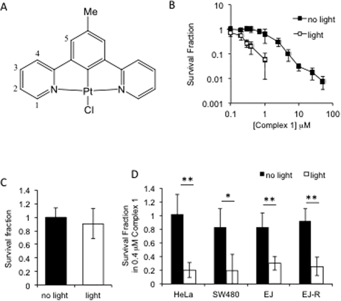
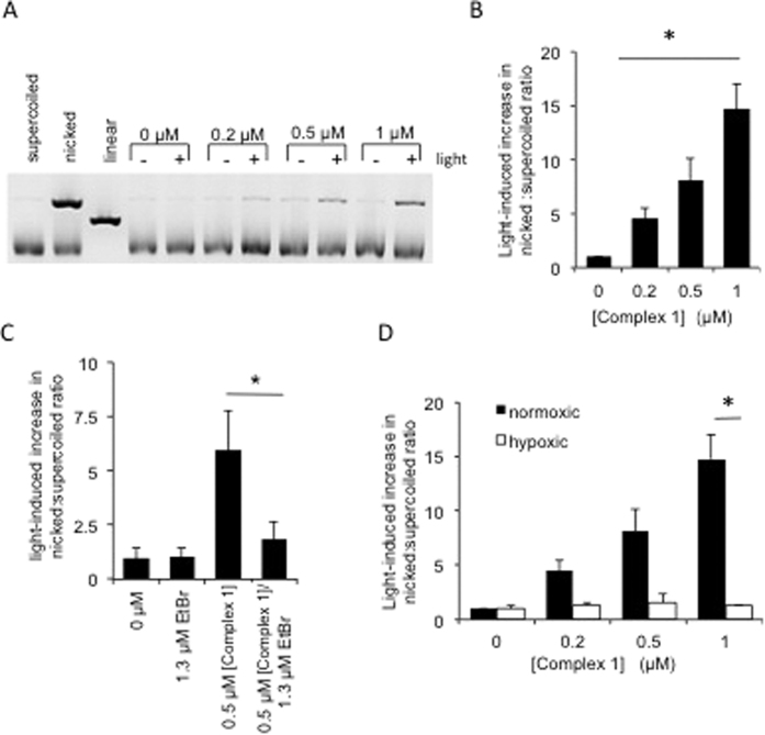
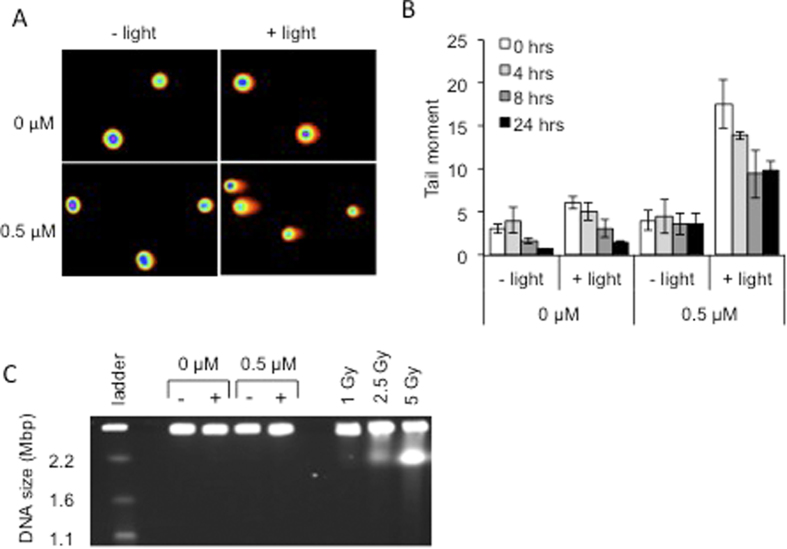
Photodynamic therapy that uses photosensitizers which only become toxic upon light-irradiation provides a strong alternative to conventional cancer treatment due to its ability to selectively target tumour material without affecting healthy tissue. Transition metal complexes are highly promising PDT agents due to intense visible light absorption, yet the majority are toxic even without light. This study introduces a small, photostable, charge-neutral platinum-based compound, Pt(II) 2,6-dipyrido-4-methyl-benzenechloride, complex 1, as a photosensitizer, which works under visible light. Activation of the new photosensitizer at low concentrations (0.1-1 μM) by comparatively low dose of 405 nm light (3.6 J cm(-2)) causes significant cell death of cervical, colorectal and bladder cancer cell lines, and, importantly, a cisplatin resistant cell line EJ-R. The photo-index of the complex is 8. We demonstrate that complex 1 induces irreversible DNA single strand breaks following irradiation, and that oxygen is essential for the photoinduced action. Neither light, nor compound alone led to cell death. The key advantages of the new drug include a remarkably fast accumulation time (diffusion-controlled, minutes), and photostability. This study demonstrates a highly promising new agent for photodynamic therapy, and attracts attention to photostable metal complexes as viable alternatives to conventional chemotherapeutics, such as cisplatin.


Figures

Figure 1
Figure 1. Photodynamic killing of cancer cells by complex 1.
(A) Structure of complex 1. (B) Survival fractions of HeLa cells following exposure to increasing doses of complex 1. Cells were pre-treated with complex 1 prior to 3 min exposure to 405 nm light (3.6 J cm−2). (C) Survival fractions of HeLa cells exposed to light in the absence of complex 1. (D) Survival fractions of cervical cancer (HeLa), colorectal cancer (SW480), bladder cancer (EJ) and cisplatin-resistant bladder cancer (EJ-R) cells. Cells were pre-treated with 400 nM complex 1 prior to 3 min exposure to 405 nm light. Data shown are mean and standard deviation of at least 3 independent experiments. Significance was determined using the Student’s T-test where n = 3 and p < 0.01 is indicated by **, and p < 0.05 is indicated by *. Survival fractions were calculated relative to untreated control.
Figure 2
Figure 2. Complex 1 binding to DNA.
(A) Metaphase spread of chromosomes extracted from HeLa cells that had been pre-incubated with complex 1. Emission image (left); light microscope image (right) and merged image (centre). (Bi) Emission decay of complex 1 recorded at 525 – 575 nm range as a function of concentrations of calf thymus DNA under 410 nm pulsed excitation. (Bii) Relative amplitudes of the emission decay component attributed to the unbound -▪-, intercalated -□-, and partially intercalated -⋄- [complex 1] at different DNA concentrations. –x- is a sum of the relative contribution of the two DNA-bound forms, -⋄- and -□-. (C) Competitive DNA binding of complex 1 versus EtBr.25 μM calf thymus DNA was incubated with 10 μM EtBr, followed by titration with complex 1 solution. The emission spectrum of the displaced EtBr has been measured. (Ci) Increase in relative emission intensity of free EtBr upon addition of [complex 1] to the DNA pretreated with EtBr. (Cii) Emission spectra from which the data in Ci were obtained. The inset shows the plot used to determine the binding constant, see text for details.
Figure 3
Figure 3. Light-induced activation of complex 1 induces DNA single strand breaks (DNA nicks) in plasmid DNA and is inhibited by addition of ethidium bromide or in hypoxic conditions.
(A) Electrophoresis of PUC19 plasmid DNA treated with (+) and without (−) 405 nm light (3.6 J cm−2) and between 0 μM and 1 μM of complex 1. 0.3 μg DNA was loaded from each sample. Lanes 1–3 represent control DNAs where nicks/single strand breaks were induced by rapid freeze thawing of DNA and linearization induced using a restriction endonuclease. (B) Quantification of the light-induced change in ratio of nicked to supercoiled DNA. Values represent fold increase in ratios in light exposed compared to non-exposed samples. (C) Quantification of the light-induced change in ratio of nicked to supercoiled DNA when ethidium bromide (EtBr) was included in the reaction. (D) Quantification of the light-induced change in ratio of nicked to supercoiled DNA when reactions were carried out under normoxic or under hypoxic (0.1% oxygen) conditions. Data shown are mean and standard deviation of at least 3 independent experiments. Significance was determined using the Student’s T-test where n = 3 and p < 0.05 is indicated by *.
Figure 4
Figure 4. Light-induced complex 1 activity selectively induces DNA single strand breaks within cells.
(A) Representative COMET assay images for each of the treatments. Images were obtained using the full spectrum function of the CometScore™ computer software. Cells were originally stained with SYBR Safe™ DNA gel stain. (B) Average Tail Moment (TM) in HeLa cells at various time points following treatment with 0.5 μM complex 1 with and without exposure to 405 nm light (3.6 J cm−2). The average TM was calculated using CometScore™ software, where at least 50 cells were analysed on each of 3 occasions and the standard deviation is shown. (C) Pulsed-field gel electrophoresis to visualise DSBs in cells treated with 0.5 μM complex 1 with and without exposure to 405 nm light. Cells were harvested immediately after treatment. Gamma irradiation (1 – 5 Gy) of cells was used as positive control for the induction of DSBs.

References

    1. Parkin D. M., Bray F., Ferlay J. & Pisani P. Global cancer statistics, 2002. CA: Can J Clin 55, 74–108 (2005). - PubMed
    1. Kamkaew A. & Burgess K. Double-Targeting Using a TrkC Ligand Conjugated to Dipyrrometheneboron Difluoride (BODIPY) Based Photodynamic Therapy (PDT) Agent. J Med Chem 56, 7608–7614 (2013). - PMC - PubMed
    1. Wu D. et al.. Efficacy of 2-(1-hexyloxyethyl)-2-devinyl pyropheophorbide-a in photodynamic therapy of human esophageal squamous cancer cells. Oncol Let 6, 1111–1119 (2013). - PMC - PubMed
    1. Yoon H.-E., Oh S.-H., Kim S.-A., Yoon J.-H. & Ahn S.-G. Pheophorbide a-mediated photodynamic therapy induces autophagy and apoptosis via the activation of MAPKs in human skin cancer cells. Oncol Rep 31, 137–144 (2014). - PubMed
    1. O’Connor A. E., Gallagher W. M. & Byrne A. T. Porphyrin and nonporphyrin photosensitizers in oncology: preclinical and clinical advances in photodynamic therapy. Photochem Photobiol 85, 1053–1074 (2009). - PubMed

Publication types