. 2016 Oct;27(10):3079-3092.
doi: 10.1681/ASN.2015090994. Epub 2016 Mar 3.

Proteomics of Urinary Vesicles Links Plakins and Complement to Polycystic Kidney Disease

Affiliations

Proteomics of Urinary Vesicles Links Plakins and Complement to Polycystic Kidney Disease

Mahdi Salih et al. J Am Soc Nephrol. 2016 Oct.

Abstract

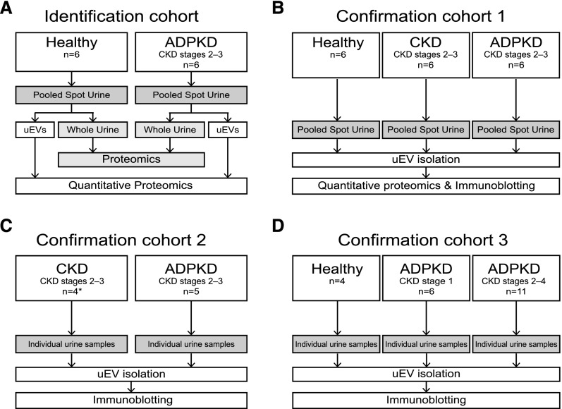
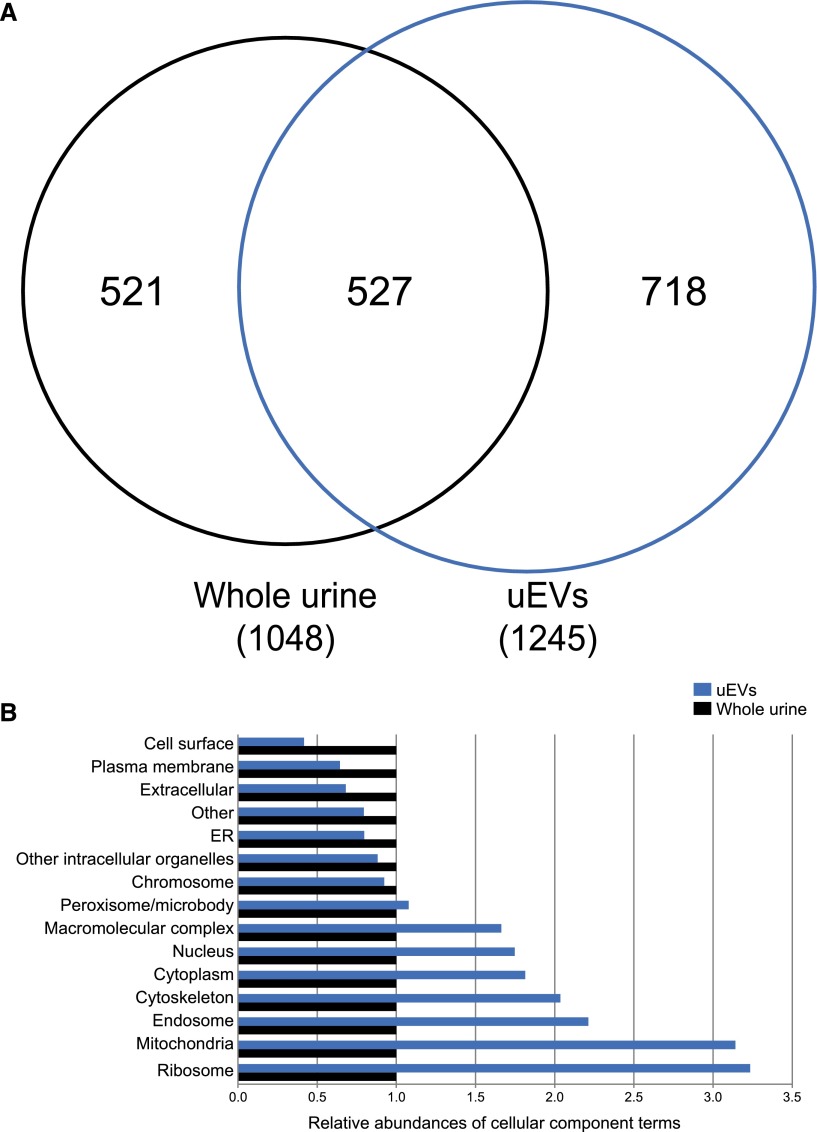
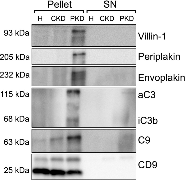
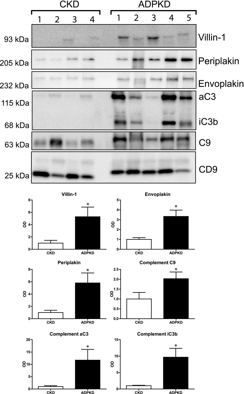
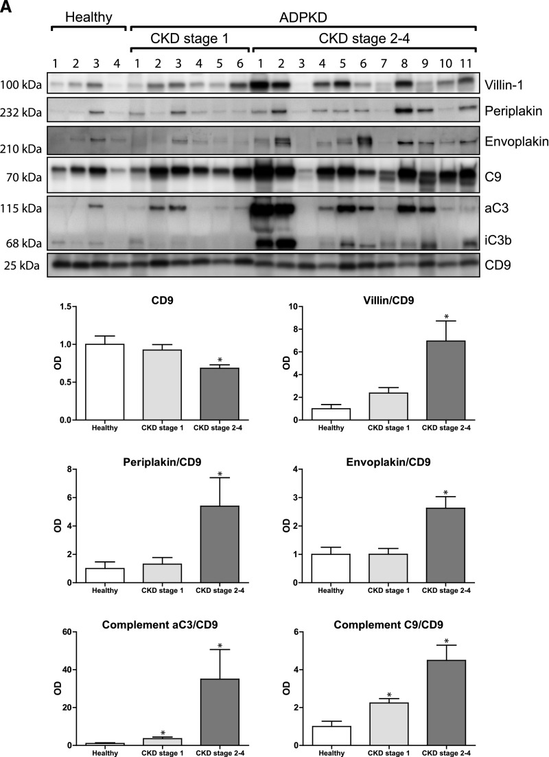
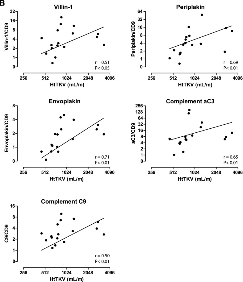
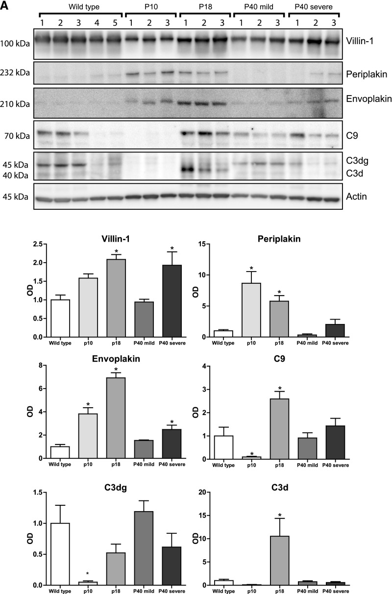
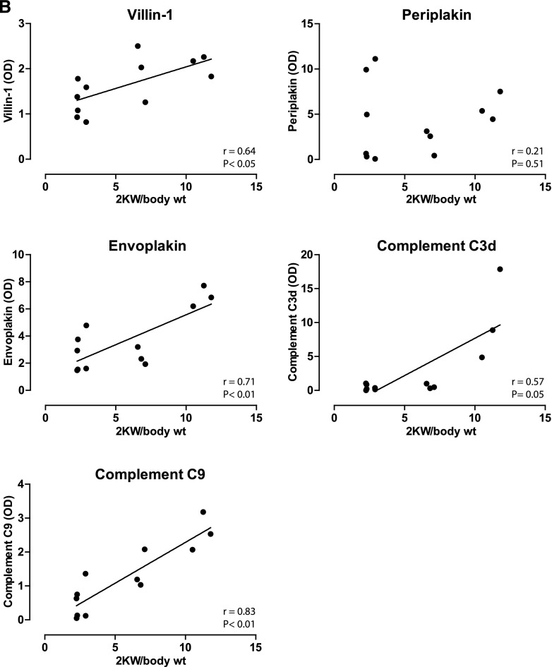
Novel therapies in autosomal dominant polycystic kidney disease (ADPKD) signal the need for markers of disease progression or response to therapy. This study aimed to identify disease-associated proteins in urinary extracellular vesicles (uEVs), which include exosomes, in patients with ADPKD. We performed quantitative proteomics on uEVs from healthy controls and patients with ADPKD using a labeled approach and then used a label-free approach with uEVs of different subjects (healthy controls versus patients with ADPKD versus patients with non-ADPKD CKD). In both experiments, 30 proteins were consistently more abundant (by two-fold or greater) in ADPKD-uEVs than in healthy- and CKD-uEVs. Of these proteins, we selected periplakin, envoplakin, villin-1, and complement C3 and C9 for confirmation because they were also significantly overrepresented in pathway analysis and were previously implicated in ADPKD pathogenesis. Immunoblotting confirmed higher abundances of the selected proteins in uEVs from three independent groups of patients with ADPKD. Whereas uEVs of young patients with ADPKD and preserved kidney function already had higher levels of complement, only uEVs of patients with advanced stages of ADPKD had increased levels of villin-1, periplakin, and envoplakin. Furthermore, all five proteins correlated positively with total kidney volume. Analysis in kidney tissue from mice with kidney-specific, tamoxifen-inducible Pkd1 deletion demonstrated higher expression in more severe stages of the disease and correlation with kidney weight for each protein of interest. In summary, proteomic analysis of uEVs identified plakins and complement as disease-associated proteins in ADPKD. These proteins are new candidates for evaluation as biomarkers or targets for therapy in ADPKD.

Keywords: ADPKD; complement; cytoskeleton; genetic renal disease.


Figures

Figure 1.
Figure 1.
Sample collection, processing, and analysis in the four study groups. (A and B) Identification and confirmation cohorts used for proteomic analysis. Quantitative proteomics was performed using dimethyl labeling in the identification cohort and label-free methods in confirmation cohort 1 (see Concise Methods). (C and D) Confirmation cohorts 2 and 3 were used for the immunoblotting analysis. *Patients from validation cohort.
Figure 2.
Figure 2.
Number of identified proteins and their cellular localization. (A) Venn diagram showing that uEVs contained a different set of unique proteins compared with whole urine. (B) Comparison of the cellular components to which the unique proteins identified whole urine (n=521) and uEVs (n=718) belong. For comparison, whole urine is set at a relative abundance of 1. ER, endoplasmic reticulum.
Figure 3.
Figure 3.
Immunoblot confirmation of five candidate proteins. Immunoblot analysis of the five selected proteins using the same pooled urine as was used for quantitative proteomics in the validation group. The first three rows show results for isolated uEVs (pellet), while the last three rows show results for the supernatant (SN), which was used as negative control. Anti-complement C3 antibody recognizes the C3 α chain (aC3) and its split product, iC3b. H, healthy persons; PKD, polycystic kidney disease.
Figure 4.
Figure 4.
Immunoblot analysis of proteins of interest in confirmation cohort 2. Immunoblot analysis comparing the proteins of interest between individual patients with CKD and ADPKD. uEVs were isolated from individual spot urine samples of patients with CKD and ADPKD (confirmation cohort 2). Anti-complement C3 antibody recognizes the C3 α chain (aC3) and its split product, iC3b. Error bars, SEM. *P<0.05.
Figure 5.
Figure 5.
Immunoblot and total kidney volume analysis of proteins of interest in confirmation cohort 3. (A) Immunoblot analysis comparing the proteins of interest in uEVs from individual healthy persons, young patients with ADPKD and preserved renal function, and patients with ADPKD and CKD stages 2–4. (B) Correlations of abundance of the uEV proteins of interest compared with height-adjusted total kidney volume (HtTKV). Spearman Rho S and P values are shown. Error bars, SEM. *P<0.05.
Figure 5.
Figure 5.
Immunoblot and total kidney volume analysis of proteins of interest in confirmation cohort 3. (A) Immunoblot analysis comparing the proteins of interest in uEVs from individual healthy persons, young patients with ADPKD and preserved renal function, and patients with ADPKD and CKD stages 2–4. (B) Correlations of abundance of the uEV proteins of interest compared with height-adjusted total kidney volume (HtTKV). Spearman Rho S and P values are shown. Error bars, SEM. *P<0.05.
Figure 6.
Figure 6.
Sucrose gradient fractionation. Sucrose gradient fractionation was performed to analyze the type of vesicles with which the identified proteins associate. Fraction 1 represents the most dense fraction. In addition to the six proteins of interest, we also analyzed CD9 and CD63 (markers for urinary exosomes) and NHE3 and AQP2 (markers for proximal tubule and collecting duct). "Pellet" refers to a part of the pooled ultracentrifugation pellet used for direct immunoblotting (positive control). Anti-complement C3 antibody recognizes the C3 α chain (aC3) and its split product, iC3b.
Figure 7.
Figure 7.
Immunoblot analysis of proteins of interest in mouse model of ADPKD. (A) Immunoblot analysis comparing the proteins of interest in kidney homogenates of three inducible ADPKD mouse models. "P" indicates the postnatal day at which the Pkd1 gene was inactivated with tamoxifen. The P40 mice were euthanized at two different time points, producing a mild (normal blood urea) or severe (elevated blood urea) phenotype. Each ADPKD group was compared with wild type. (B) Correlations between kidney weights and kidney abundances of villin-1, envoplakin, periplakin, and complements C3d and C9. Spearman Rho and P values are shown. *P<0.05. 2KW/body wt, 2 kidney weight–to–body weight ratio.
Figure 7.
Figure 7.
Immunoblot analysis of proteins of interest in mouse model of ADPKD. (A) Immunoblot analysis comparing the proteins of interest in kidney homogenates of three inducible ADPKD mouse models. "P" indicates the postnatal day at which the Pkd1 gene was inactivated with tamoxifen. The P40 mice were euthanized at two different time points, producing a mild (normal blood urea) or severe (elevated blood urea) phenotype. Each ADPKD group was compared with wild type. (B) Correlations between kidney weights and kidney abundances of villin-1, envoplakin, periplakin, and complements C3d and C9. Spearman Rho and P values are shown. *P<0.05. 2KW/body wt, 2 kidney weight–to–body weight ratio.

References

    1. Neumann HP, Jilg C, Bacher J, Nabulsi Z, Malinoc A, Hummel B, Hoffmann MM, Ortiz-Bruechle N, Glasker S, Pisarski P, Neeff H, Krämer-Guth A, Cybulla M, Hornberger M, Wilpert J, Funk L, Baumert J, Paatz D, Baumann D, Lahl M, Felten H, Hausberg M, Zerres K, Eng C Else-Kroener-Fresenius-ADPKD-Registry : Epidemiology of autosomal-dominant polycystic kidney disease: An in-depth clinical study for south-western Germany. Nephrol Dial Transplant 28: 1472–1487, 2013 - PubMed
    1. Ong AC, Devuyst O, Knebelmann B, Walz G ERA-EDTA Working Group for Inherited Kidney Diseases : Autosomal dominant polycystic kidney disease: The changing face of clinical management. Lancet 385: 1993–2002, 2015 - PubMed
    1. Gould SJ, Raposo G: As we wait: Coping with an imperfect nomenclature for extracellular vesicles. J Extracell Vesicles 2: 2, 2013 - PMC - PubMed
    1. Dear JW, Street JM, Bailey MA: Urinary exosomes: A reservoir for biomarker discovery and potential mediators of intrarenal signalling. Proteomics 13: 1572–1580, 2013 - PubMed
    1. Salih M, Zietse R, Hoorn EJ: Urinary extracellular vesicles and the kidney: Biomarkers and beyond. Am J Physiol Renal Physiol 306: F1251–F1259, 2014 - PubMed