

Small putative NANOG, SOX2, and SSEA-4-positive stem cells resembling very small embryonic-like stem cells in sections of ovarian tissue in patients with ovarian cancer
- PMID: 26940129
- PMCID: PMC4778328
- DOI: 10.1186/s13048-016-0221-3
Small putative NANOG, SOX2, and SSEA-4-positive stem cells resembling very small embryonic-like stem cells in sections of ovarian tissue in patients with ovarian cancer
Abstract
Background: In previous studies it has been found that in cell cultures of human adult ovaries there is a population of small stem cells with diameters of 2-4 μm, which are present mainly in the ovarian surface epithelium and are comparable to very small embryonic-like stem cells (VSELs) from bone marrow. These cells are not observed by histopathologists in the ovarian tissue due to their small size and unknown clinical significance. Because these cells express a degree of pluripotency, they might be involved in the manifestation of ovarian cancer. Therefore we studied the ovarian tissue sections in women with borderline ovarian cancer and serous ovarian carcinoma to perhaps identify the small putative stem cells in situ.
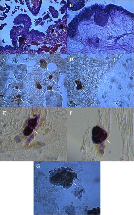
Methods: In 27 women with borderline ovarian cancer and 20 women with high-grade serous ovarian carcinoma the ovarian tissue sections were stained, per standard practice, with eosin and hematoxylin staining and on NANOG, SSEA-4 and SOX2 markers, related to pluripotency, using immunohistochemistry. We focused on the presence and localization of small putative stem cells with diameters of up to 5 μm and with the nuclei spread over nearly the full cell volume.
Results: In ovarian sections of both borderline ovarian cancer and serous ovarian carcinoma patients we were able to identify the presence of small round cells complying with the above criteria. Some of these small cells were NANOG-positive, were located among epithelial cells in the ovarian surface epithelium and as a single cell or groups of cells/clusters in typical "chambers", were found only in the presence of ovarian cancer and not in healthy ovaries and are comparable to those in fetal ovaries. We envision that these small cells could be related to NANOG-positive tumor-like structures and oocyte-like cells in similar "chambers" found in sections of cancerous ovaries, which could support their stemness and pluripotency. Further immunohistochemistry revealed a similar population of SSEA-4 and SOX2-positive cells.
Conclusions: We may conclude that putative small stem cells expressing markers, related to pluripotency, are present in the ovarian tissue sections of women with borderline ovarian cancer and high-grade serous ovarian carcinoma thus indicating their potential involvement in ovarian cancer.
Figures


Similar articles
-
Putative stem cells and epithelial-mesenchymal transition revealed in sections of ovarian tumor in patients with serous ovarian carcinoma using immunohistochemistry for vimentin and pluripotency-related markers.J Ovarian Res. 2017 Feb 23;10(1):11. doi: 10.1186/s13048-017-0306-7. J Ovarian Res. 2017. PMID: 28231820 Free PMC article.
-
Localization of the stem cell markers LGR5 and Nanog in the normal and the cancerous human ovary and their inter-relationship.Acta Histochem. 2013 May;115(4):330-8. doi: 10.1016/j.acthis.2012.09.004. Epub 2012 Oct 23. Acta Histochem. 2013. PMID: 23092806
-
The stem-cell profile of ovarian surface epithelium is reproduced in the oviductal fimbriae, with increased stem-cell marker density in distal parts of the fimbriae.Int J Gynecol Pathol. 2013 Sep;32(5):444-53. doi: 10.1097/PGP.0b013e3182800ad5. Int J Gynecol Pathol. 2013. PMID: 23896717
-
The significance of the pluripotency and cancer stem cell-related marker NANOG in diagnosis and treatment of ovarian carcinoma.Eur J Gynaecol Oncol. 2016;37(5):604-612. Eur J Gynaecol Oncol. 2016. PMID: 29786995 Review.
-
Human amnion-derived cells as a reliable source of stem cells.Curr Mol Med. 2012 Dec;12(10):1340-9. doi: 10.2174/156652412803833625. Curr Mol Med. 2012. PMID: 23016591 Review.
Cited by
-
Human amniotic epithelial cells exert anti-cancer effects through secretion of immunomodulatory small extracellular vesicles (sEV).Cancer Cell Int. 2022 Oct 29;22(1):329. doi: 10.1186/s12935-022-02755-z. Cancer Cell Int. 2022. PMID: 36307848 Free PMC article.
-
MUC1 expression is associated with ST3GAL2 and negatively correlated with the androgen receptor in castration-resistant prostate cancer.Glycoconj J. 2024 Dec;41(6):381-394. doi: 10.1007/s10719-024-10173-8. Epub 2024 Dec 24. Glycoconj J. 2024. PMID: 39718721 Free PMC article.
-
PTTG1: a Unique Regulator of Stem/Cancer Stem Cells in the Ovary and Ovarian Cancer.Stem Cell Rev Rep. 2019 Dec;15(6):866-879. doi: 10.1007/s12015-019-09911-5. Stem Cell Rev Rep. 2019. PMID: 31482269 Free PMC article.
-
Extracellular Localisation of the C-Terminus of DDX4 Confirmed by Immunocytochemistry and Fluorescence-Activated Cell Sorting.Cells. 2019 Jun 12;8(6):578. doi: 10.3390/cells8060578. Cells. 2019. PMID: 31212843 Free PMC article.
-
Do PGCCs in Solid Tumors Appear Due to Treatment-related Stress or Clonal Expansion of CSCs that Survive Oncotherapy?Stem Cell Rev Rep. 2025 Jun;21(5):1594-1606. doi: 10.1007/s12015-025-10891-y. Epub 2025 May 8. Stem Cell Rev Rep. 2025. PMID: 40338514 Review.
References
-
- Bapat SA, Mali AM, Koppikar CB, Kurrey NK. Stem and progenitor-like cells contribute to the aggressive behavior of human epithelial ovarian cencer. Cancer Res. 2005;65:3025–9. - PubMed
-
- Zhang QH, Dou HT, Xu P, Zhuang SC, Liu PS. Tumor recurrence and drug resistance properties of side population cells in high grade ovary cancer. Drug Res (Stuttg) 2015;65:153–7. - PubMed
Publication types
MeSH terms
Substances
LinkOut - more resources
Full Text Sources
Other Literature Sources
Medical
Research Materials
