. 2016 Mar 4:6:22522.
doi: 10.1038/srep22522.

Insight On Colorectal Carcinoma Infiltration by Studying Perilesional Extracellular Matrix

Affiliations

Insight On Colorectal Carcinoma Infiltration by Studying Perilesional Extracellular Matrix

Manuela Nebuloni et al. Sci Rep. .

Abstract

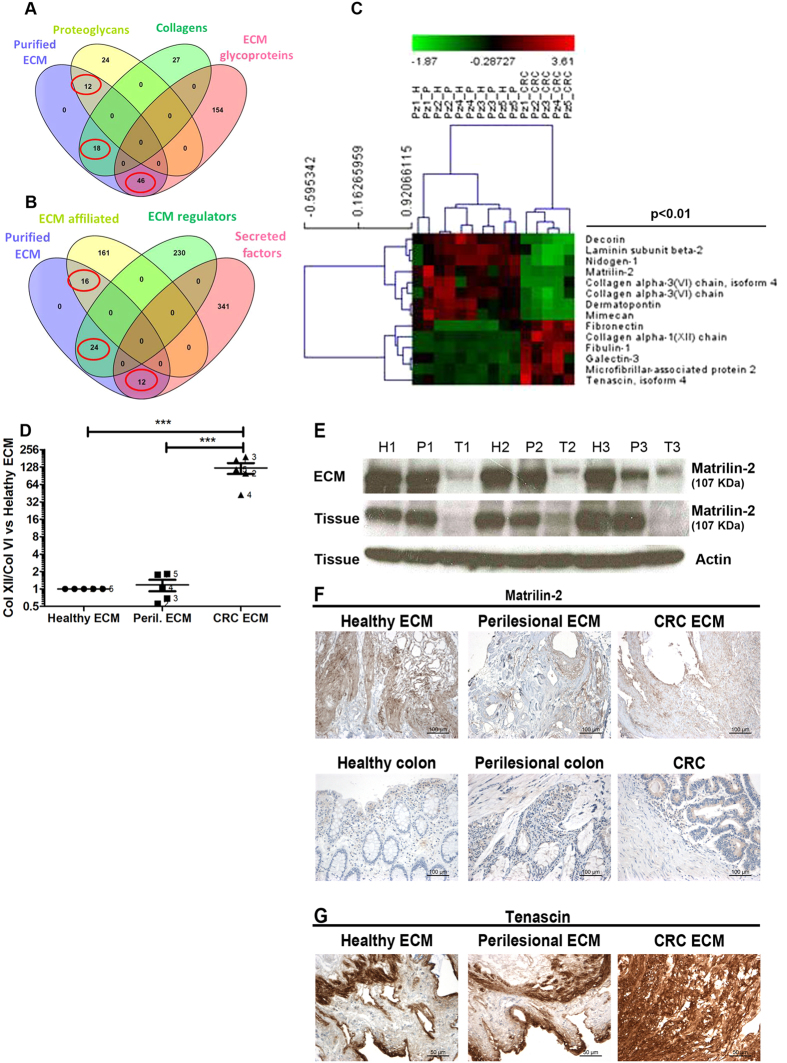
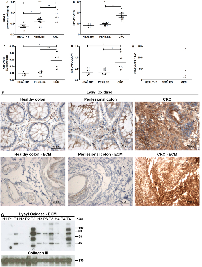
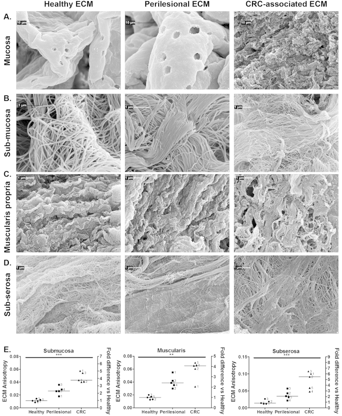
The extracellular matrix (ECM) from perilesional and colorectal carcinoma (CRC), but not healthy colon, sustains proliferation and invasion of tumor cells. We investigated the biochemical and physical diversity of ECM in pair-wised comparisons of healthy, perilesional and CRC specimens. Progressive linearization and degree of organization of fibrils was observed from healthy to perilesional and CRC ECM, and was associated with a steady increase of stiffness and collagen crosslinking. In the perilesional ECM these modifications coincided with increased vascularization, whereas in the neoplastic ECM they were associated with altered modulation of matrisome proteins, increased content of hydroxylated lysine and lysyl oxidase. This study identifies the increased stiffness and crosslinking of the perilesional ECM predisposing an environment suitable for CRC invasion as a phenomenon associated with vascularization. The increased stiffness of colon areas may represent a new predictive marker of desmoplastic region predisposing to invasion, thus offering new potential application for monitoring adenoma with invasive potential.


Figures

Figure 1
Figure 1. Tissue selection.
Pair-wised healthy colon, perilesional area and CRC were evaluated by means of hematoxylin-eosin staining (A), CD34+ blood vessels (B) and collagen (blue staining) by means of Masson Trichrome stain (C). Cr; cryptae. Lp; lamina propria. Mm; muscularis mucosae. Ca; carcinoma. Is; intratumoral stroma. *blood vessels. Pictures are representative of pair-wised tissues from one of the six patients tested and listed in Supplementary Material 1.
Figure 2
Figure 2. Protein composition of ECMs.
ECM was purified from pair-wised colon resection margins (healthy, H), perilesional areas (P) and colorectal carcinomas (CRC) from 5 patients listed in Supplementary Material 1, and proteomic analysis was performed as described in Methods. Comparison of purified ECMs (purple ellipse in the Venn’s diagrams) with the matrisome database revealed identification of 128 matrisome proteins; 76 structural components (12 proteoglycans, 18 collagens and 46 ECM glycoproteins, as highlighted by red circles in panel (A) and 52 matrix-associated components (16 ECM affiliated, 24 ECM regulators and 12 Secreted factors, as highlighted by red circles in panel (B) such as i) ECM-affiliated, as annexin, mucin, complement, calcium binding proteins S100; ii) ECM-regulators, as metallo-proteases, metallo-protease inhibitors; iii) Secreted factors bound to ECM, as latent TGF-b, growth factors, chemokines. Unsupervised hierarchical cluster analysis of LFQ intensities as derived from MaxQuant elaboration (Suppl. file 2.1) was performed upon parametric ANOVA test: the heatmap shows significantly differentially expressed ECM proteins with p-value < 0.01 (C). The ratio between collagen XII and the sum of the two forms of collagen VI reported in the above heat-map was estimated in all 5 ECMs and fold of expression was calculated vs healthy ECM (D). Western blot analysis for Matrilin-2 expression in ECMs and tissues from 3 surgical specimens; level of actin expression in tissue was used as loading control (E). Expression of Matrilin-2 was further evaluated by IHC, in pair-wised ECMs (one representative patient is shown) (F). Expression of Tenascin was evaluated by IHC (pair-wised tissues from one representative patient are shown) (G). (H) P and T in panel C and panel E indicate healthy, perilesional and tumor area, respectively. P value in panel C and asterisks in panel D indicate statistical significance.
Figure 3
Figure 3. Elastic Young’s modulus of ECM from healthy, perilesional and CRC area.
Cumulative distributions of Young’s modulus (E) values in Pa units (semilog10 scale) of the three ECMs derived from 1 representative patient (out of 3 tested and listed in the Supplementary Material 1; multi-gaussian fit representative of the cumulative distribution (solid line) and the contribution of major Gaussian modes (dotted line) were estimated (A). Median value for each ECM was estimated by weighting the relative contribution of each mode, and showed for all 3 donors (B). Mean value of Young’s modulus of healthy, perilesional and CRC ECM (C). Error bars are calculated as standard deviation of the median. Statistical significance was evaluated by means of ANOVA followed by Tukey post-test analysis. E; elasticity. Pa; pascal.
Figure 4
Figure 4. Level of collagen crosslinking, OH-Lys content of ECM proteins and lysyl oxidase in healthy, perilesional area and CRC ECMs.
Pair-wised ECMs from tissue of 5 donors (Supplementary Material 1) were evaluated for the content of collagen crosslinking HP and LP (A) and the ratio between HP/LP (B). Level of OH-lysine was estimated for all ECM proteins: sum of peak intensities, as estimated by MaxQuant software, for all OH-lysine-containing peptides was normalized to sum of ECM protein intensities in each ECM derived from 5 patients (Supplementary Material 2.7) (C). Sum of peak intensities, as estimated by MaxQuant software, for all OH-lysine-containing peptides was normalized to each protein intensity in each ECM derived from 5 patients (Supplementary Material 2.8) (D, Collagen 1A1; (E), Collagen 11A1). Asterisks indicate statistical significance evaluated by means of ANOVA. Immunohistochemistry (40× magnification) of LOX on pair-wised tissues (healthy colon, perilesional area and CRC) and the derived ECMs; data from one representative surgical sample out of 4 tested (F). Level of lysyl oxidase was evaluated by western blot using pair-wised ECMs from 4 donors (G); the molecular masses (kDa) are indicated on the right side of the panel. H, P and T in panel G indicate that ECM was isolated from pair-wised healthy, perilesional and tumor area, respectively, from 4 different surgical specimens.
Figure 5
Figure 5. Ultrastructural analysis of ECMs.
ECMs from mucosa (A), submucosa (B), muscularis propria (C) and subserosa (D) of pair-wised healthy colon, perilesional area and CRC were evaluated by means of scanning electron microscopy. Lower magnifications are reported in the Supplementary Fig. S5. Pictures show ultrastructure of pair-wised ECMs from one representative patient (#2 listed in Supplementary Material 1). Degree of organization of fibrils (anisotropy) was evaluated in pair-wised ECMs from 5 donors (E); horizontal bars indicate median value, and asterisks show statistical significance evaluated by means of Anova. Representative scanning electron micrographs used for establishing anisotropy are reported in the Supplementary Fig. S6.

References

    1. Folkman J. & Kalluri R. Cancer without disease. Nature 427, 787 (2004). - PubMed
    1. Kalluri R. & Zeisberg M. Fibroblasts in cancer. Nat Rev Cancer 6, 392–401 (2006). - PubMed
    1. Rhim A. D. et al.. Stromal elements act to restrain, rather than support, pancreatic ductal adenocarcinoma. Cancer Cell 25, 735–747 (2014). - PMC - PubMed
    1. Ozdemir B. C. et al.. Depletion of carcinoma-associated fibroblasts and fibrosis induces immunosuppression and accelerates pancreas cancer with reduced survival. Cancer Cell 25, 719–734 (2014). - PMC - PubMed
    1. Jaalouk D. E. & Lammerding J. Mechanotransduction gone awry. Nat Rev Mol Cell Biol 10, 63–73 (2009). - PMC - PubMed