Review
. 2016 Mar 8;67(9):1091-1103.
doi: 10.1016/j.jacc.2015.12.048.

Leukocytes Link Local and Systemic Inflammation in Ischemic Cardiovascular Disease: An Expanded "Cardiovascular Continuum"

Affiliations
Review

Leukocytes Link Local and Systemic Inflammation in Ischemic Cardiovascular Disease: An Expanded "Cardiovascular Continuum"

Peter Libby et al. J Am Coll Cardiol. .

Abstract

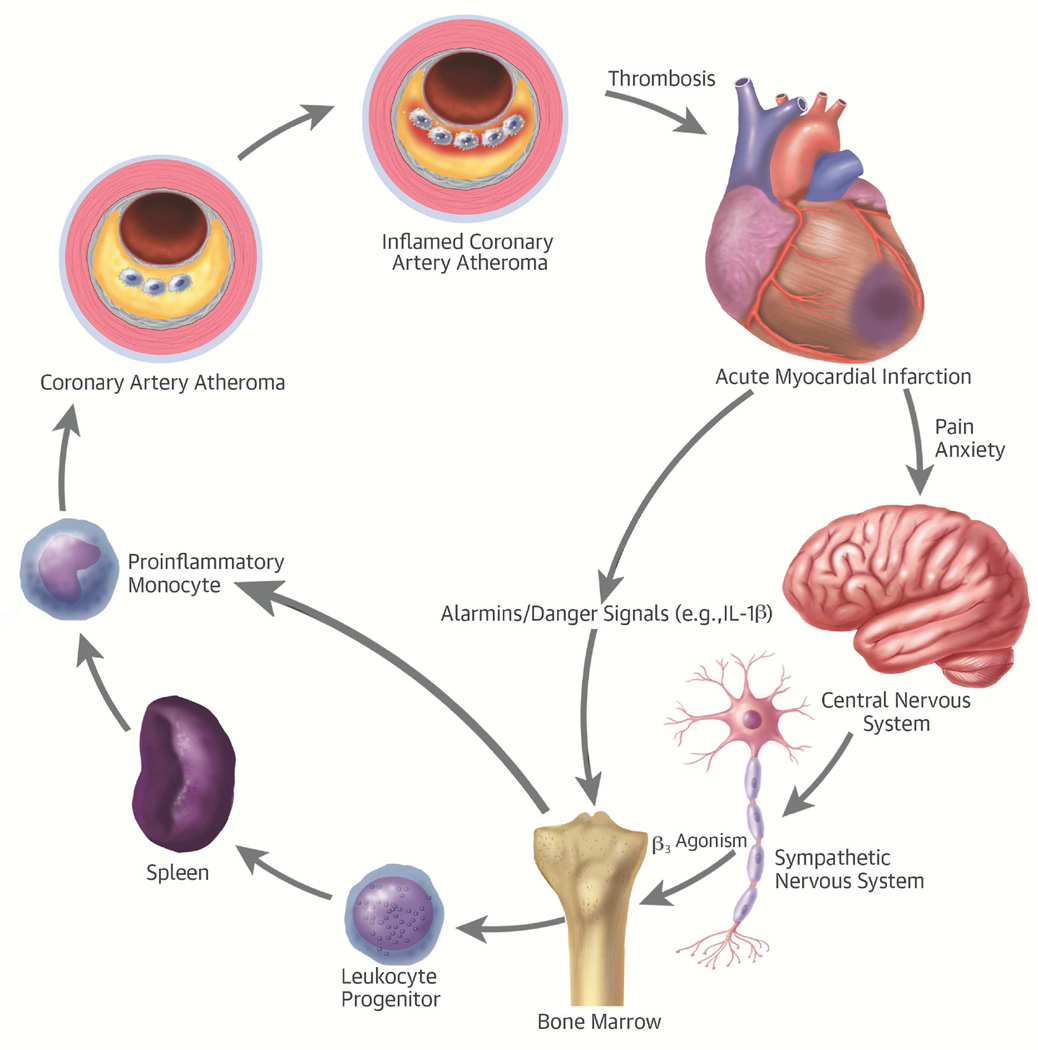
Physicians have traditionally viewed ischemic heart disease in a cardiocentric manner: plaques grow in arteries until they block blood flow, causing acute coronary and other ischemic syndromes. Recent research provides new insight into the integrative biology of inflammation as it contributes to ischemic cardiovascular disease. These results have revealed hitherto unsuspected inflammatory signaling networks at work in these disorders that link the brain, autonomic nervous system, bone marrow, and spleen to the atherosclerotic plaque and to the infarcting myocardium. A burgeoning clinical published data indicates that such inflammatory networks-far from a mere laboratory curiosity-operate in our patients and can influence aspects of ischemic cardiovascular disease that determine decisively clinical outcomes. These new findings enlarge the circle of the traditional "cardiovascular continuum" beyond the heart and vessels to include the nervous system, the spleen, and the bone marrow.

Keywords: acute coronary syndromes; myocardial infarction; white blood cells.


Figures

Figure 1
Figure 1. Heterogeneity of Mononuclear Phagocytes in the Atheroma
In early atherosclerotic plaques (shown on the left-hand side of the diagram), proinflammatory leukocytes, characterized by high levels of Ly6c and the chemokine receptor CCR2, preferentially adhere to the activated endothelial monolayer overlying the early plaque. The adherent leukocytes enter the intima by diapedesis. Accumulation of mononuclear phagocytes in the early atherosclerotic plaque occurs primarily by recruitment from blood. In the advancing atheroma (right-hand portion of the diagram), monocyte macrophage replication occurs in response to hematopoietic growth factors, some of which may arise from a specialized set of B lymphocytes known as immune response activator B cells (34). The mitotic figures indicate cellular replication. Mononuclear phagocytes resident in the intima ultimately will accumulate cholesteryl ester, forming foam cells, the hallmark of the atherosclerotic lesion. CCR = CC chemokine receptor.
Figure 2
Figure 2. Temporal Sequence and Functions of Leukocytes Localizing in the Infarcting Myocardium
Granulocytes arrive in the ischemic area within hours of the onset of infarction. The granulocytes, “first responders” to ischemic injury, elaborate pro-oxidants, proteinases, cytokines, and lipid mediators among other substances that amplify the acute inflammatory response. Examples of the mediators are shown in parentheses. In the second and third day following onset of myocardial ischemia, proinflammatory monocytes bearing high levels of the marker Ly6c and the chemokine receptor CCR2 accumulate in the infarcting tissue. These cells perform cleanup functions by secreting proteinases that can digest debris from dead or dying cells. They can phagocytose the debris and engulf dead cells by efferocytosis. These “demolition” functions can help pave the way for tissue repair. From days 4 through 7, monocytes, macrophages that exhibit reparative functions, assume predominance. In mice, these cells arise from the proinflammatory monocytes in response to transcriptional control by Nr4a1. These reparative monocytes elaborate TGF-β, which stimulates the collagen synthesis necessary for scar formation and healthy healing of the ischemic area. Angiogenic mediators, such as VEGF, stimulate microvessels characteristic of granulation tissue in the healing myocardium. These reparative monocytes can also elaborate anti-inflammatory cytokines, such as IL-10, that can quell the inflammatory response after clearance of the acutely injured cells and foster formation of a fibrous scar. IL = interleukin; MMP = matrix metalloproteinase; TGF = transforming growth factor; VEGF = vascular endothelial growth factor.
Figure 3
Figure 3. An “Echo” of Systemic Inflammation in the Atheroma
(A) The left-hand panel shows northern blots for IL-1α-(top) and IL-1β (bottom) in the aortas of nonatherosclerotic and atherosclerotic animals without or with intravenous administration of Gram-negative bacterial endotoxin (lipopolysaccharide [LPS]). Note the striking increase in mRNA encoding both isoforms of IL-1 in the atherosclerotic artery following LPS administration. (B) mRNA concentrations measured by reverse transcriptase-polymerase chain reaction in extracts of the aortas of rabbits that consumed a chow diet or graded increases in concentrations of dietary cholesterol from 0.1% to 0.9 %. Note the marked increase in IL-1β mRNA concentrations in the animals treated with intravenous endotoxin for 1 h (gray bars) versus control. These early experiments illustrate the principle that the resting atheroma can undergo inflammatory activation in response to a systemic proinflammatory stimulus. LPS = lipopolysaccharide; mRNA = messenger ribonucleic acid; RNA = ribonucleic acid. Other abbreviations as in Figure 2.
Figure 4
Figure 4. Activation of Glucose Uptake in the Arterial Wall, Spleen, and Bone Marrow in Patients With Acute Coronary Syndromes
18F-fluorodeoxyglucose (18F-FdG) uptake increased significantly in the arterial wall (aorta), bone marrow, and spleen in patients with acute coronary syndrome (ACS) versus control subjects. Reprinted from Emami et al. (67).
Figure 5
Figure 5. Regions of the Atherosclerotic Human Aorta Accumulate Imaging Reporters of Glucose Uptake (18F-FdG) and Cell Proliferation (18F-FLT)
(A) Sagittal image disclosing extensive calcification in a human aorta and carotid artery (arrows). (B) 18F-fluorodeoxyglucose (18F-FdG) and (C) 18F-fluorothymidine (18F-FLT) images show accumulation of these positron emission tomography (PET) tracers in the aortic arch (arrows). Reprinted from Ye et al. (75).
Central Illustration
Central Illustration. Leukocytes Link Local and Systemic Inflammation in Ischemic Cardiovascular Disease: The integrative pathophysiology of inflammation in ischemic heart disease
The stress of acute myocardial infarction produces an “echo” in atherosclerotic plaques. Acute myocardial infarction causes pain and anxiety that triggers sympathetic outflow from the central nervous system. β-3-adrenergic stimulation mobilizes leukocyte progenitors from their bone marrow niche. These progenitor cells can migrate to the spleen, where they can multiply in response to hematopoietic growth factors. The proinflammatory monocytes then leave the spleen and enter the atherosclerotic plaque, where they promote inflammation that can render a plaque more likely to provoke thrombosis and hence acute myocardial infarction. IL = interleukin.

Comment in

References

    1. Libby P. History of discovery: inflammation in atherosclerosis. Arterioscler Thromb Vasc Biol. 2012;32:2045–2051. - PMC - PubMed
    1. Virchow R. Cellular Pathology. London, UK: John Churchill; 1858.
    1. Virchow R. Cellular pathology. As based upon physiological and pathological histology. Lecture XVI--Atheromatous affection of arteries. 1858. Nutr Rev. 1989;47:23–25. - PubMed
    1. Nguyen AT, Gomez D, Bell RD, et al. Smooth muscle cell plasticity: fact or fiction? Circ Res. 2013;112:17–22. - PMC - PubMed
    1. Tabas I, Garcia-Cardeña G, Owens GK. Recent insights into the cellular biology of atherosclerosis. J Cell Biol. 2015;209:13–22. - PMC - PubMed

Publication types

LinkOut - more resources