. 2016 May;171(1):192-205.
doi: 10.1104/pp.15.01301. Epub 2016 Mar 3.

Defects in Peroxisomal 6-Phosphogluconate Dehydrogenase Isoform PGD2 Prevent Gametophytic Interaction in Arabidopsis thaliana

Affiliations

Defects in Peroxisomal 6-Phosphogluconate Dehydrogenase Isoform PGD2 Prevent Gametophytic Interaction in Arabidopsis thaliana

Christian Hölscher et al. Plant Physiol. 2016 May.

Abstract

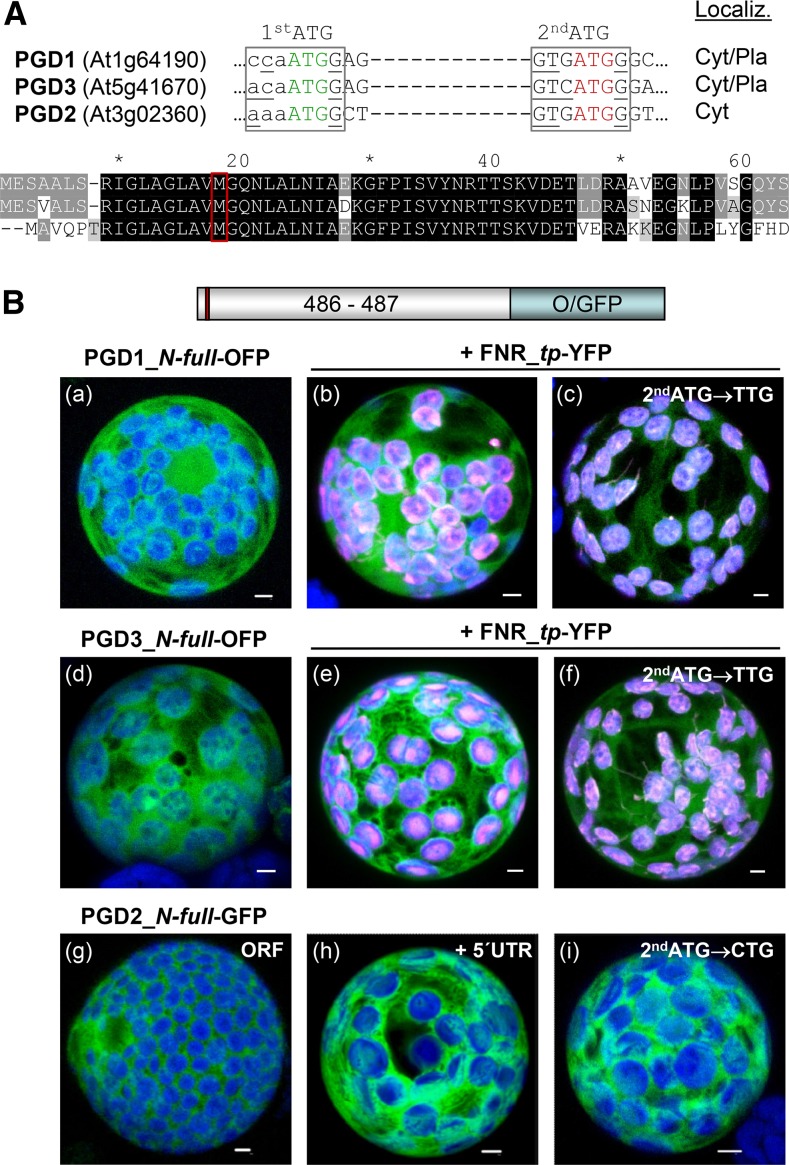
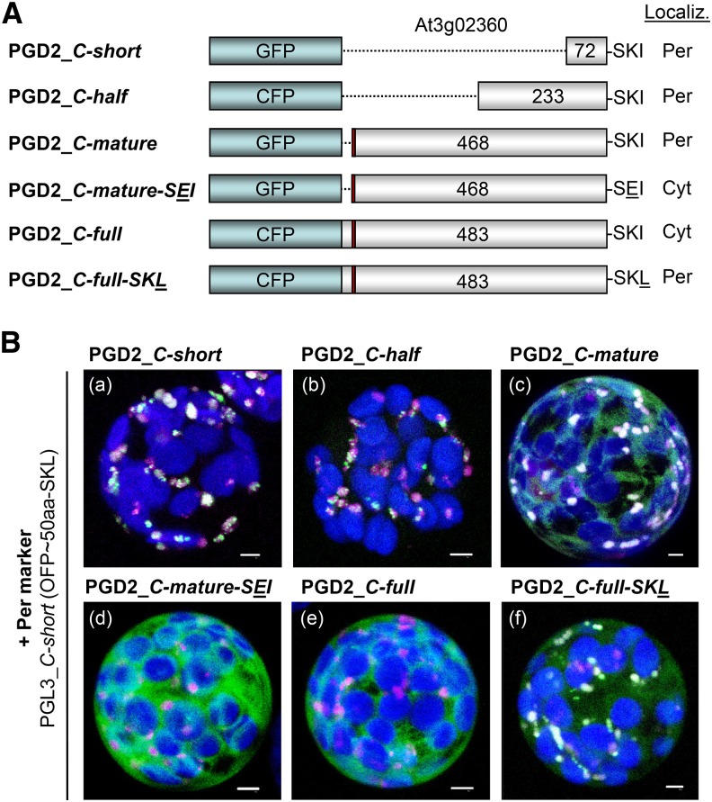
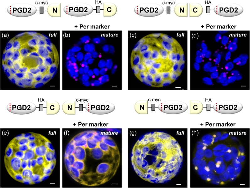
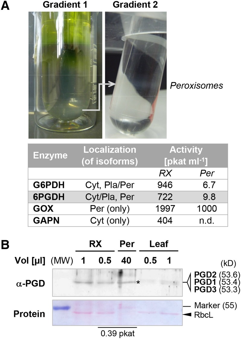
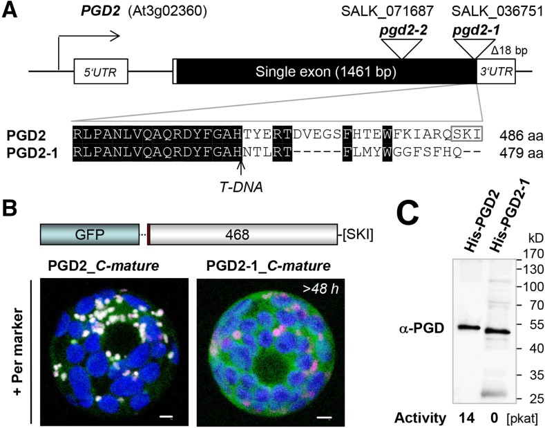
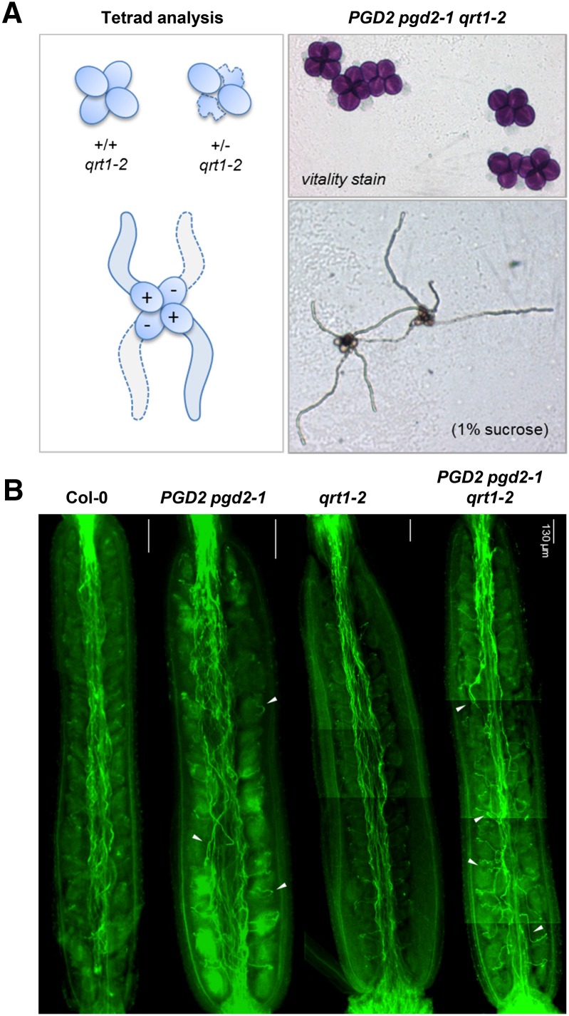
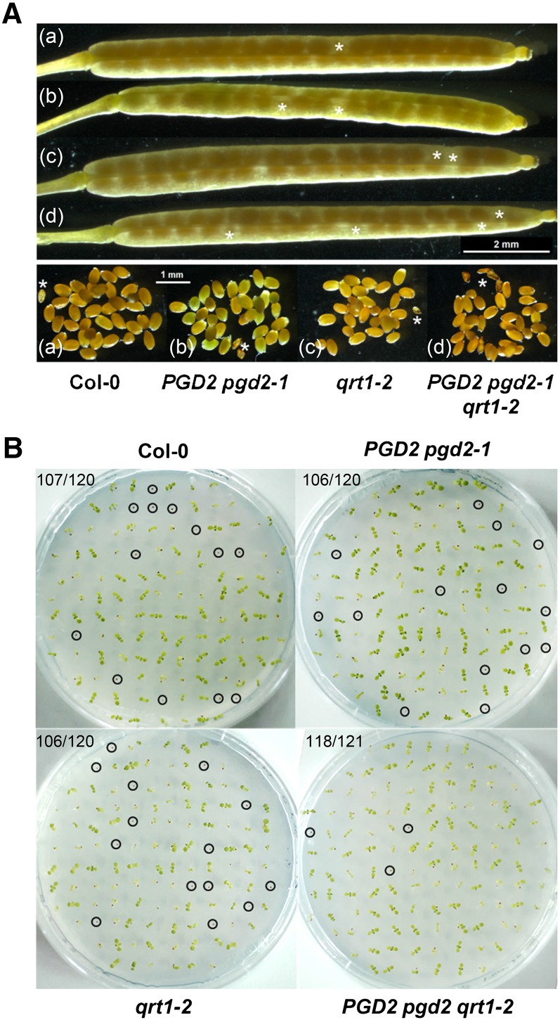
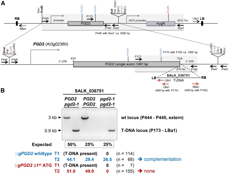
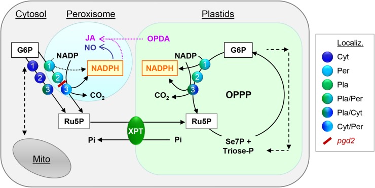
We studied the localization of 6-phosphogluconate dehydrogenase (PGD) isoforms of Arabidopsis (Arabidopsis thaliana). Similar polypeptide lengths of PGD1, PGD2, and PGD3 obscured which isoform may represent the cytosolic and/or plastidic enzyme plus whether PGD2 with a peroxisomal targeting motif also might target plastids. Reporter-fusion analyses in protoplasts revealed that, with a free N terminus, PGD1 and PGD3 accumulate in the cytosol and chloroplasts, whereas PGD2 remains in the cytosol. Mutagenesis of a conserved second ATG enhanced the plastidic localization of PGD1 and PGD3 but not PGD2. Amino-terminal deletions of PGD2 fusions with a free C terminus resulted in peroxisomal import after dimerization, and PGD2 could be immunodetected in purified peroxisomes. Repeated selfing of pgd2 transfer (T-)DNA alleles yielded no homozygous mutants, although siliques and seeds of heterozygous plants developed normally. Detailed analyses of the C-terminally truncated PGD2-1 protein showed that peroxisomal import and catalytic activity are abolished. Reciprocal backcrosses of pgd2-1 suggested that missing PGD activity in peroxisomes primarily affects the male gametophyte. Tetrad analyses in the quartet1-2 background revealed that pgd2-1 pollen is vital and in vitro germination normal, but pollen tube growth inside stylar tissues appeared less directed. Mutual gametophytic sterility was overcome by complementation with a genomic construct but not with a version lacking the first ATG. These analyses showed that peroxisomal PGD2 activity is required for guided growth of the male gametophytes and pollen tube-ovule interaction. Our report finally demonstrates an essential role of oxidative pentose-phosphate pathway reactions in peroxisomes, likely needed to sustain critical levels of nitric oxide and/or jasmonic acid, whose biosynthesis both depend on NADPH provision.


Figures

Figure 1.
Figure 1.
Localization of full-length PGD1-, PGD3-, and PGD2-reporter fusions. A, Details of the DNA sequence (top) and alignment of the N-terminal ends of the three Arabidopsis PGD isoforms (bottom). A potential second start codon (2ndATG) with Kozak consensus (bases underlined) in all isoforms is highlighted in red. Cyt/Pla, Cytosol/plastids. B, Transient expression of the indicated fusion constructs in tobacco protoplasts (48 h post transfection). PGD3 (a) and PGD1 (d) accumulate in both cytosol and chloroplasts, as confirmed by colocalization with a transit peptide fusion of ferredoxin-NADP reductase (FNR_tp-YFP; b and e). Silent mutagenesis of the second ATG in PGD1 and PGD3 results in enhanced labeling of chloroplasts (c and f). PGD2 labels the cytosol (g) as well as with the 5′ UTR (h) or mutated second ATG (i). Only maximal projections (of approximately 35 single sections) are shown (for single-channel images, see Supplemental Figs. S2 and S4). Candidate fusions are in green, chloroplast marker (FNR) is in magenta, and chlorophyll autofluorescence is in blue. Colocalization and very close signals (less than 200 nm) appear white in merged images. ORF, Open reading frame. Bars = 3 µm.
Figure 2.
Figure 2.
Analysis of reporter-PGD2 fusions. A, Schemes of the reporter-PGD2 fusions with intact or modified C-terminal ends (the position of the second ATG is marked in red) and their localization. Cyt, Cytosol; Per, peroxisomes. B, Transient expression of the indicated fusions in tobacco protoplasts (48 h post transfection). Only merged images of all channels are shown. All truncated PGD2 fusions with free C terminus localize to peroxisomes (a–c), unless PTS1 motif SKI is mutated to SEI, here shown for the mature version (d). The full-length CFP-PGD2 version remains in the cytosol (e) as EYFP-PGD2 (Reumann et al., 2007), unless PTS1 motif SKI is changed to SKL (f). Only maximal projections (of approximately 35 single sections) are shown (for single-channel images, see Supplemental Fig. S3). Candidate fusions are in green the peroxisome marker PGL3_C-short (Meyer et al., 2011) is in magenta, and chlorophyll autofluorescence is in blue. Colocalization and very close signals (less than 200 nm) appear white in merged images. Bars = 3 µm.
Figure 3.
Figure 3.
BiFC analysis of full-length versus mature PGD2 split YFP combinations in tobacco protoplasts (48 h post transfection). Independent of N- or C-terminal fusion, all full-length PGD2 versions dimerize in the cytosol (panels a,c,e,g), but are not imported by peroxisomes, even when both C-terminal ends are free (panel g). In case of the mature PGD2 versions, only the combination with two free C-terminal ends is imported by peroxisomes after dimerization in the cytosol (panel h; also in Arabidopsis protoplasts, Fig. S5), but not when only one C-terminal end is free (panel f). For two of the mature PGD2 combinations no reconstituted YFP signals were found (panels b and d). Only maximal projections (of ∼35 single sections) are shown. Candidate fusions in yellow, peroxisome marker (PGL3_C-short) in magenta, and chlorophyll autofluorescence in blue. Co-localization and very close signals (< 200 nm) appear white in merged images of all channels. Scale bars = 3 μm.
Figure 4.
Figure 4.
Purification of Arabidopsis leaf peroxisomes and immunodetection of PGD2. A, Top, Percoll/Suc (1) and Suc (2) gradients after centrifugation of 34 mL of crude extract, resulting in a band of purified peroxisomes (0.9 mL). Bottom, Table of G6PDH and 6PGDH activity, with glycolate oxidase (GOX) serving as the peroxisomal reference and nonphosphorylating GAPN as the cytosolic reference. In Arabidopsis, several isoenzymes may contribute to G6PDH (five isoforms) and 6PGDH (three isoforms) activity. Cyt, Cytosol; n.d., not detected; Pla, plastids; Per, peroxisomes; RX, crude extract. B, Immunoblot developed with the new His-PGD2 antiserum (α-PGD). Aliquots of crude extract, purified peroxisomes, and a thawed leaf extract were loaded next to each other. Crude extract and peroxisomes of similar 6PGDH activity (0.39 pkat) give comparable signals (asterisk). Together with the lack of GAPN activity (A, table), the missing band of Rubisco large subunit (RbcL) from the peroxisome fraction indicates little contribution of dually cytosolic/plastidial PGD1 and PGD3 isoforms of similar size (kD; in parentheses). MW, Molecular weight standard (PageRuler; Fermentas).
Figure 5.
Figure 5.
Subcellular localization and activity of PGD2 and C-terminally altered PGD2-1. A, Top, Scheme of the At3g02360 locus and positions of T-DNA insertions (triangles) in the single exon of PGD2. Bottom, C-terminal amino acid alignment of PGD2 and PGD2-1. The position of the T-DNA insertion is marked by the arrow, and the PTS1 motif is indicated by the gray frame. aa, Amino acids. B, Transient expression of mature PGD2 and PGD2-1 fusions in tobacco protoplasts (more than 48 h post transfection). By contrast to PGD2, PGD2-1 (with an altered C terminus) accumulates in the cytosol. Only maximal projections (of approximately 35 single sections) are shown. Candidate fusions are in green, the peroxisome (Per) marker PGL3_C-short (Meyer et al., 2011) is in magenta, and chlorophyll autofluorescence is in blue. Colocalization and very close signals (less than 200 nm) appear white in merged images. Bars = 3 µm. C, Activity test of purified His-PGD2 and His-PGD2-1 variants expressed in the G6PD-deficient E. coli strain BL21minus (Meyer et al., 2011) upon metal-chelate purification (nickel-nitrilotriacetic acid agarose [Ni-NTA] column). The immunoblot was developed with the His-PGD2 antiserum (α-PGD). 6PGDH activity (in pkat) of the elution fractions with comparable blot signal (Supplemental Fig. S6) is indicated below the blot, and apparent molecular masses (in kD) are given at right.
Figure 6.
Figure 6.
Analysis of pgd2-1 pollen tube growth in the qrt1-2 mutant. A, Tetrad analyses of PGD2 pgd2-1 in the qrt1-2 background (for details, see Supplemental Fig. S7). Alexander staining (top right) indicates the vitality of all pollen grains. Most PGD2 pgd2-1 tetrads grew four pollen tubes (like qrt1-2 alone; data not shown). B, Pollen tube growth in styles of the indicated genotypes visualized by Aniline Blue staining. Note that, compared with Col-0 (wild type) and qrt1-2, more pollen tubes of heterozygous PGD2 pgd2-1 plants grow detours (white arrowheads).
Figure 7.
Figure 7.
Seed set and germination frequency of PGD2 pgd2-1 plants. A, Mature siliques with seeds (top) produced by Col-0 wild-type (a), PGD2 pgd2-1 (b), qrt1-2 (c), and PGD2 pgd2-1 qrt1-2 (d) plants; aborted ovules are marked by white asterisks. B, Seed germination test of the indicated genotypes on Murashige and Skoog (MS) agar without Suc (day 15). Nongerminated seeds are marked by circles. A similar germination frequency indicates the absence of female defects; numbers in top left corners refer to germinated seeds versus total seeds analyzed.
Figure 8.
Figure 8.
Complementation analysis of PGD2 pgd2-1 plants after dip transformation with genomic gPGD2 constructs. A, Top, Scheme of the genomic constructs in binary vector pGSC1704 (with the NOS hygromycin B resistance cassette [HygR]). Bottom, PGD2 locus (At3g02360) with the position of the T-DNA insert (triangle) in SALK_036751 (termed pgd2-1) and the region amplified from wild-type DNA using PCR primers P446 and P447 (dashed lines) to obtain a genomic fragment for mutant complementation. Upon insertion into pBluescript SK, the genomic gPGD2 fragment with its own promoter and terminator sequences also was mutated using primers with an indicative XbaI site (Supplemental Table S1; Δ1st ATG [XbaI]). Both versions were cloned via SmaI/SalI into SnaBI/SalI sites (boldface) in the binary vector. Relevant restriction sites, promoter regions (bent closed arrows), primer-binding sites (arrows; for the T-DNA in red), as well as right (RB) and left (LB) T-DNA borders are indicated. ATG-TGA, Start-stop codons; T/Ter, terminator. B, PCR pattern obtained for the different genotypes (PGD2 and pgd2-1) upon amplification from SALK_036751 transformants harboring either wild-type (wt) gPGD2 (blue) or the version with the deleted first ATG codon (red), enforcing use of the second ATG (Fig. 1A; Supplemental Fig. S1). For primer-binding sites, see A. Fragment sizes are indicated on the left, and allele ratios (percentage) of hygromycin-resistant T1 and T2 plants are shown below (n = total number of plants analyzed). For the gPGD2 wild type, seven independent T1 lines were pooled, and for gPGD2 Δ1st ATG, three independent T1 lines were pooled.
Figure 9.
Figure 9.
The OPPP as NADPH source in plastids and peroxisomes. The scheme depicts possible compartmentation of OPPP reactions in Arabidopsis. Glucose 6-phosphate dehydrogenases (step 1) converts glucose 6-phosphate (G6P) to NADPH plus 6-phosphogluconolactone, which is used by 6-phosphogluconolactonases (step 2) to yield 6-phosphogluconate, the substrate of 6-phosphogluconate dehydrogenases (step 3). In this last step of the irreversible OPPP branch, another NADPH, ribulose 5-phosphate (Ru5P), and CO2 are produced. Ru5P (or xylulose 5-phosphate) can be exchanged for orthophosphate (Pi) via pentose-phosphate transporter XPT (Eicks et al., 2002) in the inner plastid membrane. NADPH produced by the OPPP in peroxisomes may sustain critical levels of (NO) and/or Jasmonic acid (JA). For the latter, OPDA (12-oxophytodienoic acid) has to be imported from plastids. NO and JA play important roles during fertilization and seed development. For more clarity, NADPH produced in the cytosol is not shown. Abbreviations: Cyt, Cytosolic; Per, peroxisomal; and Pla, plastidial localization. Dual targeting is indicated by mixed color symbols, the pgd2 block is high-lighted in red.

References

    1. Alexander MP. (1969) Differential staining of aborted and nonaborted pollen. Stain Technol 44: 117–122 - PubMed
    1. Alonso JM, Stepanova AN, Leisse TJ, Kim CJ, Chen H, Shinn P, Stevenson DK, Zimmerman J, Barajas P, Cheuk R, et al. (2003) Genome-wide insertional mutagenesis of Arabidopsis thaliana. Science 301: 653–657 - PubMed
    1. Bailey-Serres J, Nguyen MT (1992) Purification and characterization of cytosolic 6-phosphogluconate dehydrogenase isozymes from maize. Plant Physiol 100: 1580–1583 - PMC - PubMed
    1. Barroso JB, Corpas FJ, Carreras A, Sandalio LM, Valderrama R, Palma JM, Lupiáñez JA, del Río LA (1999) Localization of nitric-oxide synthase in plant peroxisomes. J Biol Chem 274: 36729–36733 - PubMed
    1. Boisson-Dernier A, Frietsch S, Kim TH, Dizon MB, Schroeder JI (2008) The peroxin loss-of-function mutation abstinence by mutual consent disrupts male-female gametophyte recognition. Curr Biol 18: 63–68 - PMC - PubMed

Publication types

MeSH terms