. 2016 May 1;27(9):1442-50.
doi: 10.1091/mbc.E15-12-0832. Epub 2016 Mar 3.

Subcellular optogenetic activation of Cdc42 controls local and distal signaling to drive immune cell migration

Affiliations

Subcellular optogenetic activation of Cdc42 controls local and distal signaling to drive immune cell migration

Patrick R O'Neill et al. Mol Biol Cell. .

Abstract

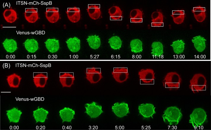
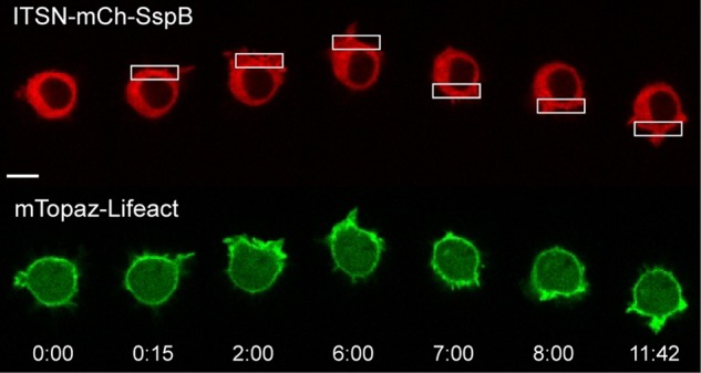
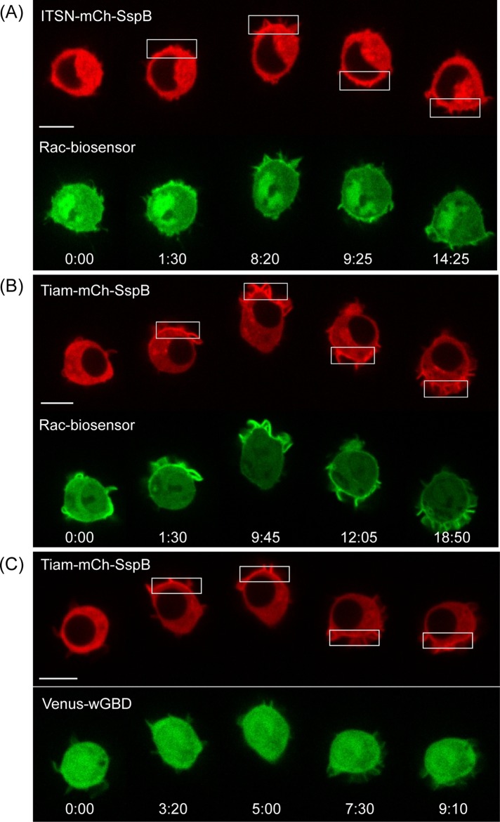
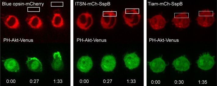
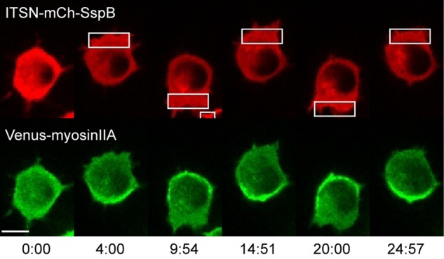
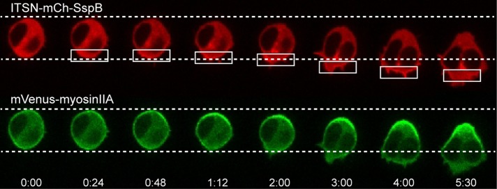
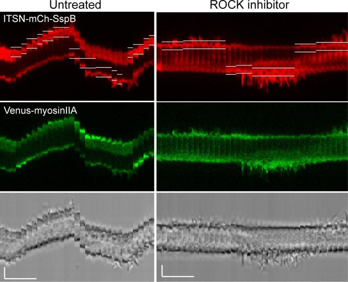
Migratory immune cells use intracellular signaling networks to generate and orient spatially polarized responses to extracellular cues. The monomeric G protein Cdc42 is believed to play an important role in controlling the polarized responses, but it has been difficult to determine directly the consequences of localized Cdc42 activation within an immune cell. Here we used subcellular optogenetics to determine how Cdc42 activation at one side of a cell affects both cell behavior and dynamic molecular responses throughout the cell. We found that localized Cdc42 activation is sufficient to generate polarized signaling and directional cell migration. The optically activated region becomes the leading edge of the cell, with Cdc42 activating Rac and generating membrane protrusions driven by the actin cytoskeleton. Cdc42 also exerts long-range effects that cause myosin accumulation at the opposite side of the cell and actomyosin-mediated retraction of the cell rear. This process requires the RhoA-activated kinase ROCK, suggesting that Cdc42 activation at one side of a cell triggers increased RhoA signaling at the opposite side. Our results demonstrate how dynamic, subcellular perturbation of an individual signaling protein can help to determine its role in controlling polarized cellular responses.


Figures

FIGURE 1:
FIGURE 1:
Chemoattractant receptors activate Cdc42 in RAW 264.7 cells. The image sequence shows a RAW cell transfected with CXCR4, Cdc42, and Venus-wGBD (Cdc42 sensor). SDF-1α was added at t = 0. Venus-wGBD selectively binds to activated Cdc42, resulting in translocation from the cytosol to the plasma membrane. The plot shows the transient decrease in cytosolic fluorescence after CXCR4 activation. Time is shown in minutes:seconds.
FIGURE 2:
FIGURE 2:
Optical stimulation of Cdc42 using a light-inducible dimerization pair. A light-inducible dimerization pair is used to achieve rapid temporal and subcellular spatial control over the membrane localization of a Cdc42 GEF (Guntas et al., 2015). The SsrA peptide is fused to the C-terminus of the Jα helix of the LOV domain. This fusion also contains a membrane-targeting domain and is referred to as iLID-CaaX. In the dark, iLID-CaaX adopts a conformation that prevents the interaction between SsrA and SspB. On optical activation, it changes conformation to make SsrA accessible for SspB binding. Optical activation is applied at one side of the cell, resulting in local recruitment of SspB to the plasma membrane. A Cdc42-selective GEF domain from ITSN is fused to SspB, so that local membrane recruitment results in local activation of Cdc42.
FIGURE 3:
FIGURE 3:
Subcellular optical activation of endogenous Cdc42 generates directional, reversible cell migration. The image sequence shows a RAW cell transfected with ITSN-tgRFPt-SspB and iLID-CaaX. The white box marks the region photoactivated with 445-nm light. Scale bar, 10 μm. Time is given in minutes:seconds. See also Supplemental Movie S1.
FIGURE 4:
FIGURE 4:
Comparison of GPCR-driven vs. Cdc42-driven migration. Optically stimulated migration of RAW cells through either localized photoactivation of GPCR signaling using blue opsin-mCherry or direct activation of Cdc42 using ITSN-tgRFPt-SspB together with iLID-CaaX. Both are capable of generating directional migration, but cells are more elongated along the direction of migration upon GPCR stimulation relative to Cdc42 activation. We found previously that asymmetric optical activation is best achieved when the optical input is positioned slightly outside of the cell using the laser power reported here (Karunarathne et al., 2013b). Here we found that asymmetric activation of the iLID construct, on the other hand, was optimized by positioning the optical input at the edge of the cell. This difference likely reflects different sensitivities of the chromophores present in opsins and LOV domains. See also Supplemental Movie S2.
FIGURE 5:
FIGURE 5:
Optically triggered membrane recruitment of ITSN generates localized Cdc42 activation. (A) Image sequence showing a RAW cell transfected with ITSN-mCh-SspB, iLID-CaaX, and Venus-wGBD. Membrane recruitment of the ITSN construct results in translocation of Venus-wGBD to the site of optical activation, demonstrating local activation of endogenous Cdc42. (B) Overexpression of wild-type Cdc42 enhances the effect. Whereas wGBD translocation in response to optical activation was only clearly detected in one of seven cells containing endogenous Cdc42 (A), it was detected in seven of nine cells when wild-type Cdc42 was cotransfected (B). Time is given in minutes:seconds. Scale bars, 10 μm. See also Supplemental Figure S4 and Supplemental Movie S3.
FIGURE 6:
FIGURE 6:
Local Cdc42 activation triggers actin polymerization at the leading edge. RAW cell transfected with ITSN-mCh-SspB, iLID-CaaX, and mTopaz-Lifeact. Optical activation of one side of the cell generates actin polymerization at the leading edge (six of six cells). Time is given in minutes:seconds. Scale bar, 10 μm. See also Supplemental Figure S5 and Supplemental Movie S4.
FIGURE 7:
FIGURE 7:
Cdc42 activation triggers Rac activation but not vice versa. RAW cells were transfected with the constructs specified in the image sequences, along with iLID-CaaX. (A) Optically triggered membrane recruitment of ITSN to activate endogenous Cdc42 resulted in local translocation of a Rac biosensor, suggesting that Rac can be activated downstream of Cdc42 in RAW cells. (B) The Tiam construct also activates Rac, consistent with Tiam’s known activity as a Rac-selective GEF. (C) The Tiam construct does not generate a detectable Cdc42 response, suggesting that Rac does not activate Cdc42 in these cells. Scale bars, 10 μm. Time is given in minutes:seconds.
FIGURE 8:
FIGURE 8:
Localized activation of Cdc42 or Rac alone does not generate increased PIP3. RAW cells transfected with the PIP3 sensor PH-Akt-Venus and blue opsin-mCherry, ITSN-mCh-SspB and iLID-CaaX, or Tiam-mCh-SspB and iLID-CaaX. Localized optical activation of the entire GPCR stimulated signaling network using blue opsin resulted in a strong PIP3 response at the front of the cell (13 of 14 cells). In contrast, localized activation of Cdc42 or Rac using the ITSN and Tiam constructs failed to generate a detectable PIP3 response (n = 11 and 12, respectively). See also Supplemental Figure S7.
FIGURE 9:
FIGURE 9:
Cdc42 activity at the leading edge induces myosin accumulation at the cell rear. RAW cell transfected with ITSN-mCh-SspB, iLID-CaaX, and Venus-myosinIIA. Optically triggered activation of Cdc42 at one side of the cell generates myosin accumulation at the opposite side. Changing the side of optical activation causes myosin to redistribute to the new cell rear. Myosin accumulation opposite the side of Cdc42 activation was observed in 56 of 63 cells. Time is given in minutes:seconds. Scale bar, 10 μm. See also Supplemental Movie S5 and Supplemental Figure S8.
FIGURE 10:
FIGURE 10:
Myosin kinetics. Cdc42-triggered changes at the cell rear occur before the generation of visible protrusions at the leading edge. A RAW cell was transfected with the constructs specified in the image sequence, together with iLID-CaaX. On localized optical activation, myosin accumulation at the cell rear can be observed before any significant protrusions occur at the cell front. In addition, retraction of the cell rear occurs before the cell front moves forward. Times are minutes:seconds.
FIGURE 11:
FIGURE 11:
Cdc42-mediated effects at both the front and back of a migrating cell involve ROCK. Kymographs showing a single RAW cell transfected with ITSN-mCh-SspB, iLID-CaaX, and Venus-myosinIIA. Localized photoactivation was performed before and after addition of 100 μM Y-27632 to the bath for 10 min to inhibit endogenous ROCK. The kymographs show that ROCK inhibition blocks myosin accumulation at the back and retraction of the cell rear. ROCK-inhibited cells also display more-pronounced membrane protrusions at the leading edge, but the cell fails to move forward. Similar results were observed for all cells tested (n = 7). White lines in the red images show the boundaries of the photoactivated regions. The brightness level has been increased for the red and green images on the right relative to those on the left in order to correct for some fluorescence decrease observed after addition of Y-27632. Scale bars, 5 μm (vertical), 5 min (horizontal). See also Supplemental Movie S6.

Similar articles

Cited by

References

    1. Allen WE, Zicha D, Ridley AJ, Jones GE. A role for Cdc42 in macrophage chemotaxis. J Cell Biol. 1998;141:1147–1157. - PMC - PubMed
    1. Arber S, Barbayannis FA, Hanser H, Schneider C, Stanyon CA, Bernard O, Caroni P. Regulation of actin dynamics through phosphorylation of cofilin by LIM-kinase. Nature. 1998;393:805–809. - PubMed
    1. Artemenko Y, Lampert TJ, Devreotes PN. Moving towards a paradigm: common mechanisms of chemotactic signaling in Dictyostelium and mammalian leukocytes. Cell Mol Life Sci. 2014;71:3711–3747. - PMC - PubMed
    1. Benard V, Bohl BP, Bokoch GM. Characterization of rac and cdc42 ­activation in chemoattractant-stimulated human neutrophils using a novel assay for active GTPases. J Biol Chem. 1999;274:13198–13204. - PubMed
    1. Benink HA, Bement WM. Concentric zones of active RhoA and Cdc42 around single cell wounds. J Cell Biol. 2005;168:429–439. - PMC - PubMed

Publication types

MeSH terms