

Cellular Barriers after Extravasation: Leukocyte Interactions with Polarized Epithelia in the Inflamed Tissue
- PMID: 26941485
- PMCID: PMC4749818
- DOI: 10.1155/2016/7650260
Cellular Barriers after Extravasation: Leukocyte Interactions with Polarized Epithelia in the Inflamed Tissue
Abstract
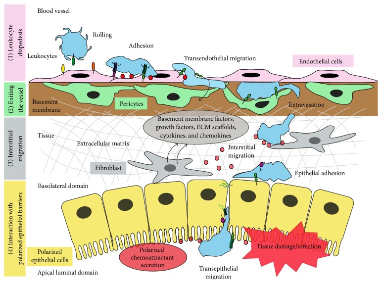
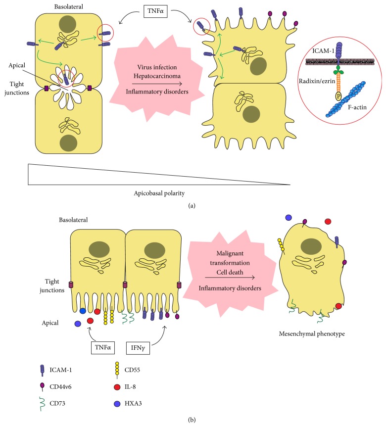
During the inflammatory response, immune cells egress from the circulation and follow a chemotactic and haptotactic gradient within the tissue, interacting with matrix components in the stroma and with parenchymal cells, which guide them towards the sites of inflammation. Polarized epithelial cells compartmentalize tissue cavities and are often exposed to inflammatory challenges such as toxics or infections in non-lymphoid tissues. Apicobasal polarity is critical to the specialized functions of these epithelia. Indeed, a common feature of epithelial dysfunction is the loss of polarity. Here we review evidence showing that apicobasal polarity regulates the inflammatory response: various polarized epithelia asymmetrically secrete chemotactic mediators and polarize adhesion receptors that dictate the route of leukocyte migration within the parenchyma. We also discuss recent findings showing that the loss of apicobasal polarity increases leukocyte adhesion to epithelial cells and the consequences that this could have for the inflammatory response towards damaged, infected or transformed epithelial cells.
Figures
Similar articles
-
Apicobasal polarity controls lymphocyte adhesion to hepatic epithelial cells.Cell Rep. 2014 Sep 25;8(6):1879-1893. doi: 10.1016/j.celrep.2014.08.007. Epub 2014 Sep 18. Cell Rep. 2014. PMID: 25242329
-
Functional insights on the polarized redistribution of leukocyte integrins and their ligands during leukocyte migration and immune interactions.Immunol Rev. 2007 Aug;218:147-64. doi: 10.1111/j.1600-065X.2007.00529.x. Immunol Rev. 2007. PMID: 17624951 Review.
-
The leading edge during dorsal closure as a model for epithelial plasticity: Pak is required for recruitment of the Scribble complex and septate junction formation.Development. 2010 Jun;137(12):2023-32. doi: 10.1242/dev.045088. Development. 2010. PMID: 20501591
-
Topographically grooved gel inserts for aligning epithelial cells during air-liquid-interface culture.Biomater Sci. 2015 Jan;3(1):121-33. doi: 10.1039/c4bm00237g. Epub 2014 Sep 9. Biomater Sci. 2015. PMID: 26214196
-
How cells tell up from down and stick together to construct multicellular tissues - interplay between apicobasal polarity and cell-cell adhesion.J Cell Sci. 2021 Nov 1;134(21):jcs248757. doi: 10.1242/jcs.248757. Epub 2021 Oct 29. J Cell Sci. 2021. PMID: 34714332 Free PMC article. Review.
Cited by
-
Evaluation of Placentation and the Role of the Aryl Hydrocarbon Receptor Pathway in a Rat Model of Dioxin Exposure.Environ Health Perspect. 2021 Nov;129(11):117001. doi: 10.1289/EHP9256. Epub 2021 Nov 8. Environ Health Perspect. 2021. PMID: 34747641 Free PMC article.
-
Viable Allogeneic Mitochondria Transplantation Improves Gas Exchange and Alveolar-Capillary Permeability in Rats with Endotoxin-Induced Acute Lung Injuries.Int J Med Sci. 2022 Jun 6;19(6):1036-1046. doi: 10.7150/ijms.73151. eCollection 2022. Int J Med Sci. 2022. PMID: 35813297 Free PMC article.
-
Pneumolysin Induces 12-Lipoxygenase-Dependent Neutrophil Migration during Streptococcus pneumoniae Infection.J Immunol. 2020 Jan 1;204(1):101-111. doi: 10.4049/jimmunol.1800748. Epub 2019 Nov 27. J Immunol. 2020. PMID: 31776202 Free PMC article.
-
ICAM-1 nanoclusters regulate hepatic epithelial cell polarity by leukocyte adhesion-independent control of apical actomyosin.Elife. 2024 Apr 10;12:RP89261. doi: 10.7554/eLife.89261. Elife. 2024. PMID: 38597186 Free PMC article.
-
Targeted transcriptional profiling of the tumor microenvironment reveals lymphocyte exclusion and vascular dysfunction in metastatic osteosarcoma.Oncoimmunology. 2019 Jun 27;8(9):e1629779. doi: 10.1080/2162402X.2019.1629779. eCollection 2019. Oncoimmunology. 2019. PMID: 31428529 Free PMC article.
References
Publication types
MeSH terms
LinkOut - more resources
Full Text Sources
Other Literature Sources
