

Viral Outbreak in Corals Associated with an In Situ Bleaching Event: Atypical Herpes-Like Viruses and a New Megavirus Infecting Symbiodinium
- PMID: 26941712
- PMCID: PMC4761846
- DOI: 10.3389/fmicb.2016.00127
Viral Outbreak in Corals Associated with an In Situ Bleaching Event: Atypical Herpes-Like Viruses and a New Megavirus Infecting Symbiodinium
Abstract
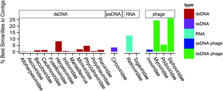
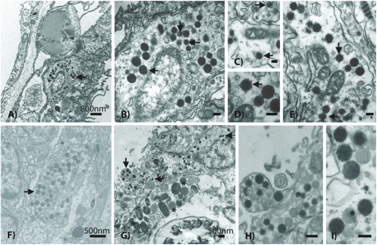
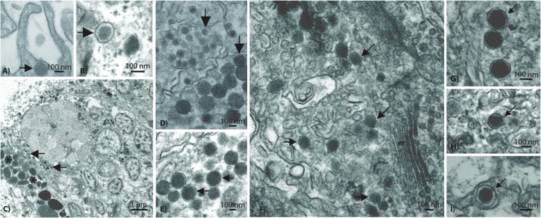
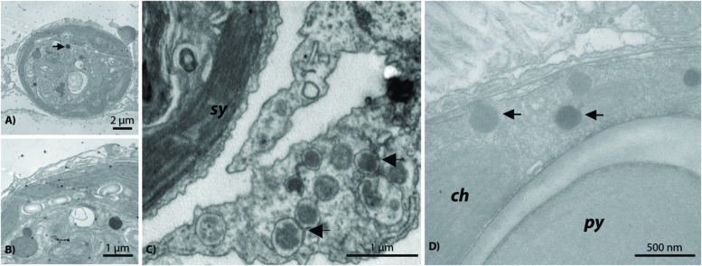
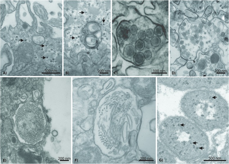
Previous studies of coral viruses have employed either microscopy or metagenomics, but few have attempted to comprehensively link the presence of a virus-like particle (VLP) to a genomic sequence. We conducted transmission electron microscopy imaging and virome analysis in tandem to characterize the most conspicuous viral types found within the dominant Pacific reef-building coral genus Acropora. Collections for this study inadvertently captured what we interpret as a natural outbreak of viral infection driven by aerial exposure of the reef flat coincident with heavy rainfall and concomitant mass bleaching. All experimental corals in this study had high titers of viral particles. Three of the dominant VLPs identified were observed in all tissue layers and budding out from the epidermis, including viruses that were ∼70, ∼120, and ∼150 nm in diameter; these VLPs all contained electron dense cores. These morphological traits are reminiscent of retroviruses, herpesviruses, and nucleocytoplasmic large DNA viruses (NCLDVs), respectively. Some 300-500 nm megavirus-like VLPs also were observed within and associated with dinoflagellate algal endosymbiont (Symbiodinium) cells. Abundant sequence similarities to a gammaretrovirus, herpesviruses, and members of the NCLDVs, based on a virome generated from five Acropora aspera colonies, corroborated these morphology-based identifications. Additionally sequence similarities to two diagnostic genes, a MutS and (based on re-annotation of sequences from another study) a DNA polymerase B gene, most closely resembled Pyramimonas orientalis virus, demonstrating the association of a cosmopolitan megavirus with Symbiodinium. We also identified several other virus-like particles in host tissues, along with sequences phylogenetically similar to circoviruses, phages, and filamentous viruses. This study suggests that viral outbreaks may be a common but previously undocumented component of natural bleaching events, particularly following repeated episodes of multiple environmental stressors.
Keywords: herpesvirus; megavirus; nucleocytoplasmic large DNA virus (NCLDV); tropical coral reef; virome; virus-like particle (VLP).
Figures
Similar articles
-
Unique nucleocytoplasmic dsDNA and +ssRNA viruses are associated with the dinoflagellate endosymbionts of corals.ISME J. 2013 Jan;7(1):13-27. doi: 10.1038/ismej.2012.75. Epub 2012 Jul 12. ISME J. 2013. PMID: 22791238 Free PMC article.
-
Potential role of viruses in white plague coral disease.ISME J. 2014 Feb;8(2):271-83. doi: 10.1038/ismej.2013.137. Epub 2013 Aug 15. ISME J. 2014. PMID: 23949663 Free PMC article.
-
Coral-associated viral communities show high levels of diversity and host auxiliary functions.PeerJ. 2017 Nov 17;5:e4054. doi: 10.7717/peerj.4054. eCollection 2017. PeerJ. 2017. PMID: 29158985 Free PMC article.
-
The Occurrence of Mixed Infections of Symbiodinium (Dinoflagellata) within Individual Hosts.J Phycol. 2012 Dec;48(6):1306-16. doi: 10.1111/j.1529-8817.2012.01220.x. Epub 2012 Sep 17. J Phycol. 2012. PMID: 27009983 Review.
-
Metagenomic characterization of viral communities in corals: mining biological signal from methodological noise.Environ Microbiol. 2015 Oct;17(10):3440-9. doi: 10.1111/1462-2920.12803. Epub 2015 Mar 27. Environ Microbiol. 2015. PMID: 25708646 Review.
Cited by
-
Influence of Chemotaxis and Swimming Patterns on the Virulence of the Coral Pathogen Vibrio coralliilyticus.J Bacteriol. 2018 Jul 10;200(15):e00791-17. doi: 10.1128/JB.00791-17. Print 2018 Aug 1. J Bacteriol. 2018. PMID: 29555697 Free PMC article.
-
Engineering Strategies to Decode and Enhance the Genomes of Coral Symbionts.Front Microbiol. 2017 Jun 30;8:1220. doi: 10.3389/fmicb.2017.01220. eCollection 2017. Front Microbiol. 2017. PMID: 28713348 Free PMC article.
-
Revisiting the rules of life for viruses of microorganisms.Nat Rev Microbiol. 2021 Aug;19(8):501-513. doi: 10.1038/s41579-021-00530-x. Epub 2021 Mar 24. Nat Rev Microbiol. 2021. PMID: 33762712 Review.
-
Corallivory and the microbial debacle in two branching scleractinians.ISME J. 2018 Apr;12(4):1109-1126. doi: 10.1038/s41396-017-0033-5. Epub 2018 Jan 16. ISME J. 2018. PMID: 29339825 Free PMC article.
-
Association of coral algal symbionts with a diverse viral community responsive to heat shock.BMC Microbiol. 2017 Aug 17;17(1):174. doi: 10.1186/s12866-017-1084-5. BMC Microbiol. 2017. PMID: 28818037 Free PMC article.
References
-
- Baker A. C., Cunning R. (2015). “Coral “bleaching” as a generalized stress response to environmental disturbance,” in Diseases of Coral, eds Woodley C. M., Downs C. A., Bruckner A. W., Porter J. W., Galloway S. B. (Hoboken, NJ: John Wiley & Sons, Inc.), 396–409.
-
- Bettarel Y., Thuy N. T., Huy T. Q., Hoang P. K., Bouvier T. (2013). Observation of virus-like particles in thin sections of the bleaching scleractinian coral Acropora cytherea. J. Mar. Biol. Assoc. 93 909–912. 10.1017/S0025315411002062 - DOI
LinkOut - more resources
Full Text Sources
Other Literature Sources