. 2016 Feb 23:7:192.
doi: 10.3389/fpls.2016.00192. eCollection 2016.

Arabidopsis thaliana: A Model Host Plant to Study Plant-Pathogen Interaction Using Rice False Smut Isolates of Ustilaginoidea virens

Affiliations

Arabidopsis thaliana: A Model Host Plant to Study Plant-Pathogen Interaction Using Rice False Smut Isolates of Ustilaginoidea virens

Mebeaselassie Andargie et al. Front Plant Sci. .

Abstract

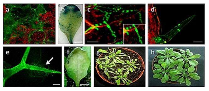
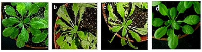
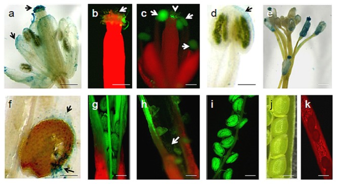
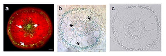
Rice false smut fungus which is a biotrophic fungal pathogen causes an important rice disease and brings a severe damage where rice is cultivated. We established a new fungal-plant pathosystem where Ustilaginoidea virens was able to interact compatibly with the model plant Arabidopsis thaliana. Disease symptoms were apparent on the leaves of the plants after 6 days of post inoculation in the form of chlorosis. Cytological studies showed that U. virens caused a heavy infestation inside the cells of the chlorotic tissues. Development and colonization of aerial mycelia in association with floral organ, particularly on anther and stigma of the flowers after 3 weeks of post inoculation was evident which finally caused infection on the developing seeds and pod tissues. The fungus adopts a uniquely biotrophic infection strategy in roots and spreads without causing a loss of host cell viability. We have also demonstrated that U. virens isolates infect Arabidopsis and the plant subsequently activates different defense response mechanisms which are witnessed by the expression of pathogenesis-related genes, PR-1, PR-2, PR-5, PDF1.1, and PDF1.2. The established A. thaliana-U. virens pathosystem will now permit various follow-up molecular genetics and gene expression experiments to be performed to identify the defense signals and responses that restrict fungal hyphae colonization in planta and also provide initial evidence for tissue-adapted fungal infection strategies.

Keywords: Arabidopsis thaliana; PR genes; Ustilaginoidea virens; endophytic colonization; plant defensin gene; plant–pathogen interaction.


Figures

FIGURE 1
FIGURE 1
Infection of 4-week-old Arabidopsis thaliana plants (ecotype Columbia) with Ustilaginoidea virens. (a) Germinating conidia on the surface of the leaf; (b) GUS stained Arabidopsis leaf after 72 hpi; (c) a branched mycelium proliferating on the leaf after 72 hpi. The inset shows magnification of the section; (d) and (e) spores and hypha attached on trichomes; (f) formation of chlorotic lesion at the site of inoculation of U. virens isolates after 96 hpi; (g) Infected Arabidopsis plants after 3 dpi; (h) Control Arabidopsis plants. Bars = 100 μm (a,d,e), 20 μm (c) and 50 μm (b,f).
FIGURE 2
FIGURE 2
Progression of disease symptoms caused by U. virens infection. The disease symptoms in Arabidopsis ecotype, Col-0: (a) 0 dpi; (b) 3 dpi; (c) 6 dpi; (d) Control.
FIGURE 3
FIGURE 3
U. virens-Arabidopsis flower interaction. (a) Visible intense GUS histochemical staining on different floral regions of infected Arabidopsis flower by U. virens 3 weeks after post-inoculation; (b) GFP tagged U. virens hyphae growing on the stigma 3 weeks after post-inoculation; (c) GFP-tagged aerial mycelia on the male and female parts of Arabidopsis flower 3 weeks after post-inoculation; (d) U. virens infected male floral structure showing the GUS stain; (e) Colonization of GUS labeled aerial mycelia on the stem, anther, and stigma of the flowers after 3 weeks of post inoculation; (f–i) Mycelium development on siliques and seeds as well as shriveled pod formation from infection of flower tissue 28 days after inoculation with GFP and GUS labeled and transformed strains of U. virens; (j) Control showing Arabidopsis siliques and seeds; (k) Uninfected Arabidopsis pod under epifluorescence microscopy. Bars = 40 μm (g–j), 20 μm (a–d,f,k), 150 μm (e).
FIGURE 4
FIGURE 4
Early colonization stages of Arabidopsis roots by U. virens. (a) hyphae growing along the epidermis parallel to the longitudinal axis of the root and forming lateral hyphopodia (arrow), 2 dpi. The inset shows magnification of the upper section of the root; (b) younger GUS stained hyphae extensively colonize the root surface, forming a network around the root; (c) heavy colonization of the Arabidopsis root by U. virens after 10 dpi; (d) root cells become colonized by hyphae in a mosaic pattern, leaving some cells uninfected after 10 dpi; (e) root of the uninfected Arabidopsis plant under bright field microscopy (Control); (f) uninfected Arabidopsis root under epifluorescence microscopy. Bars = 100 μm (a,c,d), 40 μm (e,f) and 50 μm (b).
FIGURE 5
FIGURE 5
Advanced colonization stages of Arabidopsis root by U. virens expressing GFP and GUS histochemical staining. (a) cortical cells are colonized by GFP-tagged hyphae (arrow) after 21 dpi; (b) semi-thin sectioning of Arabidopsis root showing a heavy colonization of epidermis, cortex and the endodermis after 21 dpi by the GUS labeled hyphae; (c) Semi-thin sectioning of an uninfected 21-day-old Arabidopsis root under bright field microscopy (Control). Bars = 100 μm (a) and 20 μm (b,c).
FIGURE 6
FIGURE 6
Real-time RT-PCR analysis of PR1, PR2, PR5, PDF1.1, and PDF1.2 gene expression in Arabidopsis leaves (A), roots (B), flowers (C), and pods (D) upon a treatment with U. virens. Arabidopsis leaves and roots were inoculated by adding a suspension of germinated microconidia in order to reach the concentration of 5 × 10⁵ conidia mL-1. Control leaves, flowers, roots, and pods were treated with sterile distilled water. Expression levels were normalized with respect to the housekeeping gene Actin. Data bars represent the mean ± SD of three repeats of the transcripts assessed at 0, 24, 48, 72, and 96 h post-inoculation for leaves and roots while flowers and pod gene transcripts were assessed 5 weeks post inoculation.

References

    1. Abdellatif L., Bouzid S., Kaminskyj S., Vujanovic V. (2009). Endophytic hyphal compartmentalization is required for successful symbiotic Ascomycota association with root cells. Mycol. Res. 113 782–791. 10.1016/j.mycres.2009.02.013 - DOI - PubMed
    1. Asano Y., Hiramoto T., Nishino R., Aiba Y., Kimura T., Yoshihara K., et al. (2012). Critical role of gut microbiota in the production of biologically active, free catecholamines in the gut lumen of mice. Am. J. Physiol. Gastrointest. Liver Physiol. 303 G1288–G1295. 10.1152/ajpgi.00341.2012 - DOI - PubMed
    1. Bohman S., Staal J., Thomma B. P., Wang M., Dixelius C. (2004). Characterisation of an Arabidopsis-Leptosphaeria maculans pathosystem: resistance partially requires camalexin biosynthesis and is independent of salicylic acid, ethylene and jasmonic acid signalling. Plant J. 37 9–20. 10.1046/j.1365-313X.2003.01927.x - DOI - PubMed
    1. Calo L., García I., Gotor C., Romero L. C. (2006). Leaf hairs influence phytopathogenic fungus infection and confer an increased resistance when expressing a Trichoderma α-13-glucanase. J. Exp. Bot. 57 3911–3920. 10.1093/jxb/erl155 - DOI - PubMed
    1. Chamberlain A. C. (1975). “The movement of plant particles in plant communities,” in Vegetation and the Atmosphere ed. Monteith J. L. (New York, NY: Academic Press; ) 155–204.

LinkOut - more resources