

Histopathological Study of Cyclosporine Pulmonary Toxicity in Rats
- PMID: 26941796
- PMCID: PMC4749835
- DOI: 10.1155/2016/2973274
Histopathological Study of Cyclosporine Pulmonary Toxicity in Rats
Abstract
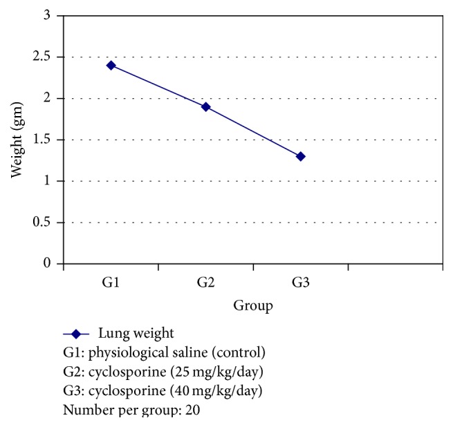
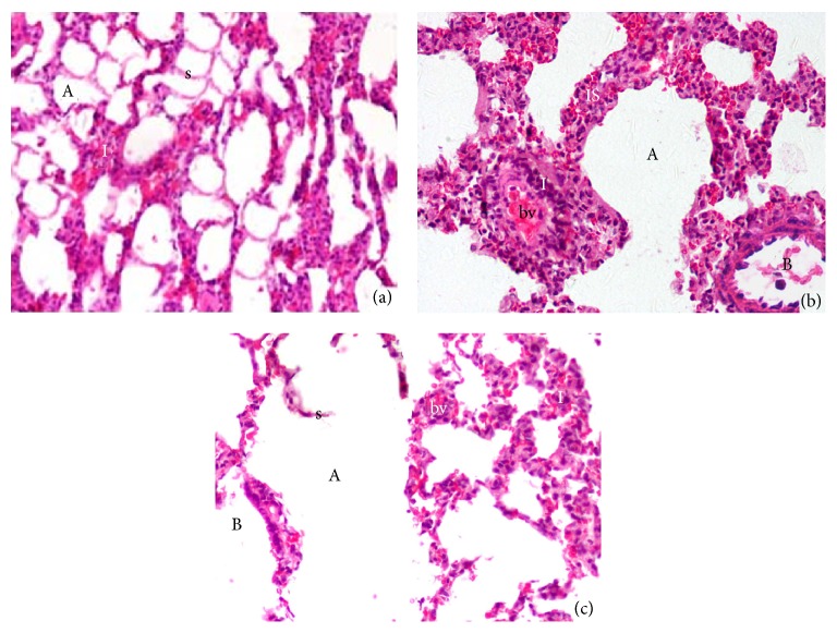
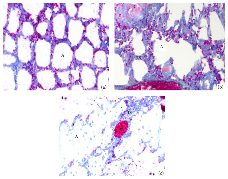
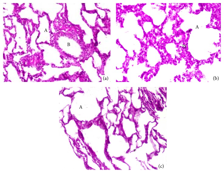
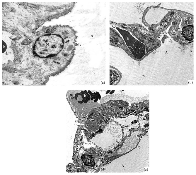
Cyclosporine is considered one of the common worldwide immunosuppressive drugs that are used for allograft rejection prevention. However, articles that address adverse effects of cyclosporine use on the vital organs such as lung are still few. This study aims to investigate pulmonary toxic effect of cyclosporine in rats by assessment of pulmonary histopathological changes using light and electron microscope examination. Sixty male adult albino rats were divided into three groups; each group consists of twenty rats. The first received physiological saline while the second and third groups received 25 and 40 mg/kg/day of cyclosporine, respectively, by gastric gavage for forty-five days. Cyclosporine reduced the lung and body weight with shrinkage or pyknotic nucleus of pneumocyte type II, degeneration of alveoli and interalveolar septum beside microvilli on the alveolar surface, emphysema, inflammatory cellular infiltration, pulmonary blood vessels congestion, and increase of fibrous tissues in the interstitial tissues and around alveoli with negative Periodic Acid-Schiff staining. Prolonged use of cyclosporine induced pulmonary ultrastructural and histopathological changes with the lung and body weight reduction depending on its dose.
Figures
Similar articles
-
Leflunomide prolongs pulmonary allograft and xenograft survival.J Heart Lung Transplant. 1995 Nov-Dec;14(6 Pt 1):1136-44. J Heart Lung Transplant. 1995. PMID: 8719461
-
Protective effect of ellagic acid against cyclosporine A-induced histopathological, ultrastructural changes, oxidative stress, and cytogenotoxicity in albino rats.Ultrastruct Pathol. 2016 Jul-Aug;40(4):205-21. doi: 10.1080/01913123.2016.1203854. Epub 2016 Jul 18. Ultrastruct Pathol. 2016. PMID: 27430433
-
[Expression and mechanism of osteoactivin in the kidney of SD rats after acute cyclosporine A toxicity].Zhong Nan Da Xue Xue Bao Yi Xue Ban. 2011 Sep;36(9):881-8. doi: 10.3969/j.issn.1672-7347.2011.09.012. Zhong Nan Da Xue Xue Bao Yi Xue Ban. 2011. PMID: 21946207 Chinese.
-
Histopathological and biochemical changes in lung tissues of rats following administration of fluoride over several generations.J Appl Toxicol. 2003 Nov-Dec;23(6):437-46. doi: 10.1002/jat.935. J Appl Toxicol. 2003. PMID: 14635268
-
Toxicology and carcinogenesis studies of androstenedione (CAS No. 63-05-8) in F344/N rats and B6C3F1 mice (gavage studies).Natl Toxicol Program Tech Rep Ser. 2010 Sep;(560):1, 7-31,33-171 passim. Natl Toxicol Program Tech Rep Ser. 2010. PMID: 21037592 Review.
Cited by
-
Optical-Spectrometry-Based Method for Immunosuppressant Medicine Level Detection in Aqueous Solutions.Sensors (Basel). 2018 Jun 22;18(7):2001. doi: 10.3390/s18072001. Sensors (Basel). 2018. PMID: 29932121 Free PMC article.
References
-
- Rezzani R. Exploring cyclosporine A-side effects and the protective role-played by antioxidants: the morphological and immunohistochemical studies. Histology and Histopathology. 2006;21(1–3):301–316. - PubMed
LinkOut - more resources
Full Text Sources
Other Literature Sources
