. 2016 Feb 13;6(4):533-44.
doi: 10.7150/thno.14315. eCollection 2016.

Mifepristone Suppresses Basal Triple-Negative Breast Cancer Stem Cells by Down-regulating KLF5 Expression

Affiliations

Mifepristone Suppresses Basal Triple-Negative Breast Cancer Stem Cells by Down-regulating KLF5 Expression

Rong Liu et al. Theranostics. .

Abstract

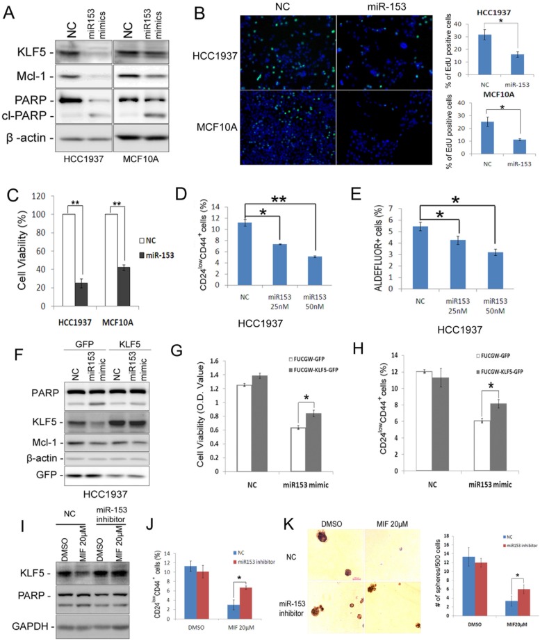
Triple-negative breast cancer (TNBC) is currently the most malignant subtype of breast cancers without effective targeted therapies. Mifepristone (MIF), a drug regularly used for abortion, has been reported to have anti-tumor activity in multiple hormone-dependent cancers, including luminal type breast cancers. In this study, we showed that MIF suppressed tumor growth of the TNBC cell lines and patient-derived xenografts in NOD-SCID mice. Furthermore, MIF reduced the TNBC cancer stem cell (CSC) population through down-regulating KLF5 expression, a stem cell transcription factor over-expressed in basal type TNBC and promoting cell proliferation, survival and stemness. Interestingly, MIF suppresses the expression of KLF5 through inducing the expression of miR-153. Consistently, miR-153 decreases CSC and miR-153 inhibitor rescued MIF-induced down-regulation of the KLF5 protein level and CSC ratio. Taken together, our findings suggest that MIF inhibits basal TNBC via the miR-153/KLF5 axis and MIF may be used for the treatment of TNBC.

Keywords: Cancer Stem Cell; KLF5; Mifepristone; Triple-negative Breast Cancer; miR-153..


Conflict of interest statement

Competing Interests: Authors have no conflicts of interest.

Figures

Figure 1
Figure 1
MIF suppresses triple-negative breast cell growth and induces apoptosis in vivo and in vitro. A. MIF suppressed HCC1937 tumor growth in NOD SCID mice in a dosage dependent manner. HCC1937 cells were injected into the fat pat of female NOD SCID mice. When the average tumor size reached about 50 mm3 after inoculation, the mice were randomly and equally distributed into three groups (n=6/group): placebo, 30 mg MIF pellet (60 days release), and 60 mg MIF pellet (60 days release). Tumor size were measured weekly for 7 weeks. B. MIF significantly decreased tumor weights compared to the placebo group (p<0.001, t-test) in a dosage dependent manner. Tumors were collected 7 weeks after MIF implantation. C. MIF did not affect the body weight of mice. The mice were weighted at the end of the experiment.D. MIF suppressed MC1 PDX growth in vivo. MC1 cells were injected into the fat pat of female NOD SCID mice. Tumor size was measured twice per week once tumors became palpable. Twelve mice carried MC1 xenografts were randomly distributed into two groups equally when the tumor size reached around 50 mm3 and were given 1 mg MIF (dissolved in tea oil) or vehicle control per day by i.p. for 25 days.E. MIF significantly decreased tumor weights compared to the vehicle control (p<0.05, t-test). Tumors were collected 25 days after MIF treatment.F. MIF did not affect the body weight of mice. The mice were weighted at the end of the experiment. G. MIF significantly inhibited DNA synthesis in both HCC1937 and MCF10A by using the Click-iTTM EdU Alexa Fluor® 647 Imaging Kit. The quantitative results are shown in the right graphs. *P<0.05; **P<0.01, t-test.H. MIF down-regulated the KLF5 protein levels and induced the cleavage of PARP and caspase 3 in a dosage-dependent manner in both HCC1937 and MCF10A cells. Protein levels were detected by WB. β-actin was used as the loading control.
Figure 2
Figure 2
MIF decreases CSC of HCC1937 and PDX cells. A. The percentage of CD24low/CD44+ cells was assessed using the flow cytometry analysis. HCC1937 cells were treated with MIF for 24 h at indicated concentration. **, P<0.01, t-test.B. MIF suppresses sphere formation and CSC self-renewal in HCC1937 cells. HCC1937 cells were treated with 10 or 20μM MIF or vehicle control for 24 h. The cells were stained with typan blue and counted to eliminate dead cells before seeding in ultra-low attachment dishes for sphere culture. 14 days later, mammospheres were collected and dissociated for passaging and the 2nd round of sphere culture. Data are shown as averages ± SD. **, P<0.01, t-test.C&D. The percentage of lin-CD24low/CD44+ cells was assessed using flow cytometry. MC1 and UM1 PDX derived cells were treated with MIF for 24 h at indicated dosage or applied for sphere culture together with MIF or vehicle control. *, P<0.05; **, P<0.01, t-test.E. MIF suppressed UM1 PDX growth in vivo. UM1 cells were injected into the fat pat of female NOD-SCID mice. Mice carried UM1 xenografts were randomly distributed into two groups equally when the tumor size reached around 50 mm3 and were given 1 mg MIF (dissolved in tea oil) or vehicle control per day by i.p. *, P<0.05, t-test. F. UM1 xenografts collected from F were dissociated, and the percentage of lin-CD24low/CD44+ cells was analyzed.G. Dissociated UM1 cells from MIF or control group were submitted to sphere culture in ultra-low attachment dishes for 2 weeks. *, P<0.05, t-test.H. Dissociated UM1 xenograft cells were injected to the fat pad of nude mice at indicated numbers, 5 weeks later, xenografts were collected for incidence analysis.
Figure 3
Figure 3
MIF suppresses KLF5 expression in time and dosage-dependent manners in basal breast epithelial cells and KLF5 depletion decreases CSC. A. The KLF5 protein level was suppressed by MIF in time- and dosage-dependent manners in MCF10A and HCC1937. The cells were treated with MIF for 24 h at indicated concentration or at 40 μM for indicated time. β-actin was used as the loading control. B. KLF5 is highly expressed in CSC population. The CSC-enriched CD24low/CD44+ population and the non-CSC CD24+/CD44- population were sorted for WB analysis. GAPDH was used as loading control. C. KLF5 protein level was significantly silenced by three different shRNAs in HCC1937. D. Stable knockdown of KLF5 by different shRNAs decreased the percentage of CD24low/CD44+ CSC in HCC1937. *, P<0.05, t-test. E. Stable depletion of KLF5 in HCC1937 by different shRNAs significantly suppressed mammosphere formation compared to the Lucsh control. The quantitative results are shown in the right graph. *, P<0.05, t-test.
Figure 4
Figure 4
Ectopic over-expression of KLF5 partially rescues MIF-induced apoptosis and CSC reduction in HCC1937. A. KLF5 over-expression decreases MIF-induced PARP cleavage in HCC1937. HCC1937 cell were transiently electroporated with pBabe-KLF5 or pBabe vector control, followed by treating with 40 μM MIF for 24 h. The apoptosis marker cl-PARP was detected by WB. B. Over-expression of KLF5 significantly rescued the MIF-induced CD24low/CD44+ CSC reduction in HCC1937. The quantitative results are shown in the right graph. *, P<0.05, t-test.C. Ectopic expression of KLF5 in HCC1937 partially rescued the MIF-induced mammosphere formation reduction. Mammospheres were counted 12 days after seeding. *, P<0.05, t-test.
Figure 5
Figure 5
MIF suppresses the expression of KLF5 through inducing the miR-153 expression. A. MIF increased the expression of miR-21, miR-152 and miR-153 in both HCC1937 and MCF10A cells. U6 was used as the internal control. B. miR-153 mimics (50 nM) decreased the KLF5 protein level in HCC1937 and MCF10A cells. The cells were transfected with miRNA mimics for two days. KLF5 protein levels were detected by WB. C. miR-153 suppressed the KLF5 3'-UTR luciferase reporter activity. HEK293FT cells were transfected with miR-153 mimics (50 nM) and pMIR-KLF5 3'-UTR or pMIR-KLF5 3'-UTRm reporter together with pCMV-renilla control. Two days after transfection, the cells were collected for the dual-luciferase reporter assay. **, P<0.001, t-test. D. miR-153 inhibitor rescued the MIF-induced KLF5 protein decrease in MCF10A cells. The cells were transfected with either miR-153 inhibitor or negative control miRNA inhibitor. One day after transfection, the cells were treated with 20 μM MIF for 24 h. E. The miR-153 inhibitor rescued the MIF-induced KLF5 protein decrease in HCC1937 cells.
Figure 6
Figure 6
miR-153 suppresses basal breast epithelial cell proliferation, survival and CSC. A. miR-153 suppressed the expression of KLF5 and Mcl-1 and induced the cleavage of PARP in HCC1937 and MCF10A cells. The cells were transfected with 50 nM negative control (NC) or miR-153 mimics for two days. B. miR-153 inhibits DNA synthesis using the EdU assay in HCC1937 and MCF10A cells. The quantitative results are shown in the right graphs.C. The cell viability was significantly reduced by miR-153 in HCC1937 and MCF10A cells. The cells were plated in 24-well plate at a density of 5×10⁴ in triplicates. One day after plating, the cells were transfected with 50 nM NC or miR-153 mimics. Two days after microRNA transfection, the cells were fixed for SRB assays. **, P<0.01, t-test.D&E. miR-153 suppressed the percentage of CD24low/CD44+ (D) and ALDH+ (E) cells in HCC1937 in a dosage dependent manner. The cells were transfected with 50 nM NC or miR-153 mimics for 2 days. *, P<0.05; **, P<0.001, t-test.F. Ectopic over-expression of KLF5 decreases MIF-induced PARP cleavage in HCC1937. HCC1937 cell were infected with FUCGW-KLF5 or FUCGW vector control, followed by treating with 20 μM MIF for 24 h. The apoptosis marker cl-PARP was detected by WB. β-actin was used as loading control.G. FUCGW-KLF5 or vector control lentivirus-infected HCC1937 cells were plated in 48-well plate at 3×10⁴/well and transfected with miR-153 mimic or negative control for 2 days. Cell viability was analyzed using SRB assays.H. FUCGW-KLF5 or vector control lentivirus-infected HCC1937 cells were transfected with miR-153 mimic or negative control for 2 days before being subjected for analyzing the percentage of CD24low/CD44+ populations. I-K. miR-153 inhibitor or negative control transfected cells were treated with MIF or vehicle control for 24 h. The cells were collected for WB (I), CD24low/CD44+ populations analysis (J) or mammosphere culture for 2 weeks (K). *, P<0.05, t-test.

References

    1. Desmedt C, Ruiz-Garcia E, Andre F. Gene expression predictors in breast cancer: current status, limitations and perspectives. Eur J Cancer. 2008;44:2714–20. - PubMed
    1. Goldhirsch A, Wood WC, Coates AS. et al. Strategies for subtypes-dealing with the diversity of breast cancer: highlights of the St. Gallen International Expert Consensus on the Primary Therapy of Early Breast Cancer 2011. Annals of oncology: official journal of the European Society for Medical Oncology / ESMO. 2011;22:1736–47. - PMC - PubMed
    1. Dent R, Trudeau M, Pritchard KI. et al. Triple-negative breast cancer: clinical features and patterns of recurrence. Clinical cancer research: an official journal of the American Association for Cancer Research. 2007;13:4429–34. - PubMed
    1. Rakha EA, El-Sayed ME, Green AR. et al. Prognostic markers in triple-negative breast cancer. Cancer. 2007;109:25–32. - PubMed
    1. Lehmann BD, Bauer JA, Chen X. et al. Identification of human triple-negative breast cancer subtypes and preclinical models for selection of targeted therapies. The Journal of clinical investigation. 2011;121:2750–67. - PMC - PubMed

Publication types

MeSH terms