

Focal hepatic lesions characterisation by different sonographic techniques: a prospective analysis
- PMID: 26941878
- PMCID: PMC4762844
- DOI: 10.1007/s40477-015-0172-3
Focal hepatic lesions characterisation by different sonographic techniques: a prospective analysis
Abstract
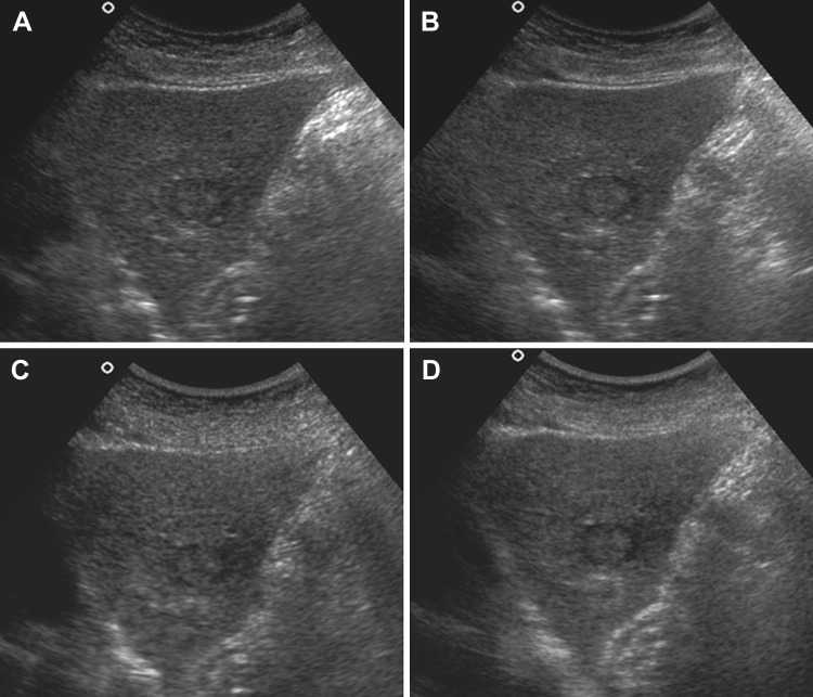
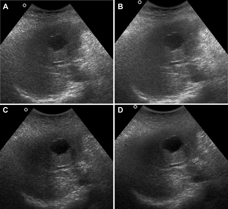
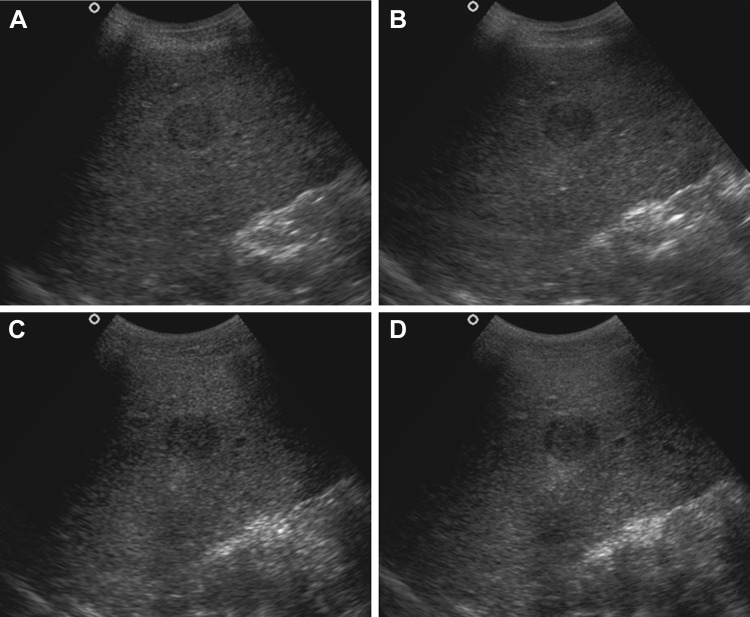
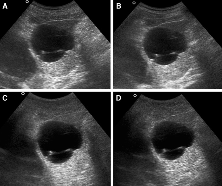
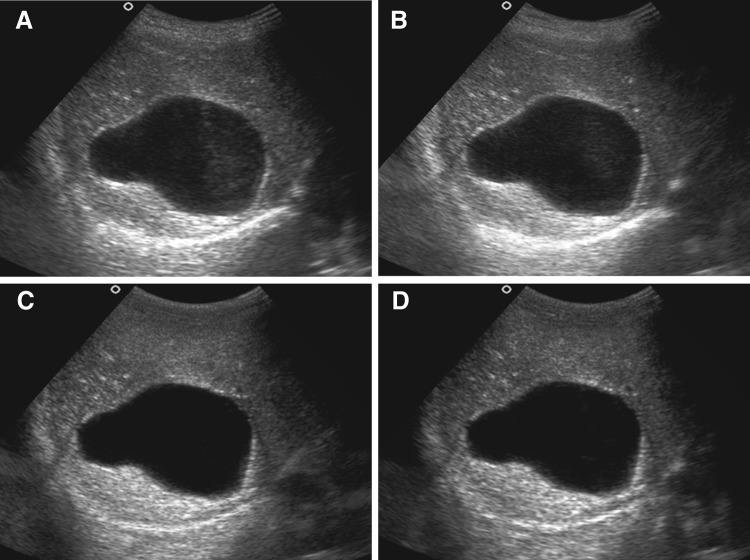
Introduction: Ultrasound is usually the first diagnostic investigation for the assessment of liver lesions. Apart from conventional sonography (CS), new grey-scale sonographic techniques have been developed which have increased the application of ultrasound in liver imaging. The present study was undertaken to compare image quality of CS, real-time compound sonography (RTCS), tissue harmonic sonography (THS) and tissue harmonic compound sonography (THCS) in focal liver lesions.
Materials and methods: 100 patients with focal hepatic lesions were enroled. Lesions were divided into solid and cystic group. Solid lesions were evaluated for lesion conspicuity and elimination of artefacts. For cystic lesions, lesion conspicuity, posterior acoustic enhancement and internal echoes within the lesion were evaluated. Grading was done using 3-5-point scales. Overall image quality was assessed depending on the total points.
Results: 78 solid and 22 cystic liver lesions were included. THCS showed superior results for lesion conspicuity, elimination of artefacts and overall image quality in solid lesions. RTCS showed similar results as THCS for lesion conspicuity and overall image quality in solid lesions. THS gave better results in cystic lesions for all imaging parameters. Results of THCS though slightly inferior, showed no significant difference from THS, in cystic lesions. CS was found to have least diagnostic value in characterisation.
Conclusions: For evaluation of focal hepatic lesions, a combination of compound and harmonic sonography, i.e. THCS, is the preferred sonographic technique.
Introduzione: l’ecografia è di solito l’indagine radiologica iniziale, non invasiva e semplice, per la valutazione delle lesioni epatiche. Oltre alla tradizionale ecografia in scala di grigi (CS), sono state sviluppate delle nuove tecniche, che hanno aumentato le applicazioni dell’ecografia nell’imaging del fegato. Il presente studio è stato intrapreso per confrontare la qualità delle immagini in: CS, ecografia in tempo reale con compound (RTCS), armonica tissutale (THS) e armonica tissutale con compound (THC) nello studio delle lesioni focali epatiche.
Materiali e metodi: sono stati arruolati 100 pazienti con lesioni focali epatiche. Le lesioni sono state divise in solide e cistiche. Per le lesioni solide sono stati valutati la visibilità e l’eliminazione di artefatti. Per le lesioni cistica sono stati valutati la visibilità, il rinforzo acustico posteriore e gli echi all’interno delle lesioni. La classificazione è stata effettuata utilizzando una scala da 3 a 5. La qualità generale dell’immagine è stata valutata in base al totale dei punti.
Risultati: sono state incluse 78 lesioni epatiche solide e 22 cistiche. La THC ha mostrato risultati superiori per la visibilità delle lesioni, l’eliminazione di artefatti e la qualità complessiva dell’immagine delle lesioni solide. La RTCS ha mostrato risultati simili a quelli di THC per la visibilità delle lesioni e la qualità complessiva dell’immagine delle lesioni solide. THS ha dato risultati migliori per tutti i parametri di imaging delle lesioni cistiche. I risultati della THC, anche se leggermente inferiori, non hanno mostrato differenze significativa dalla THS, nelle lesioni cistiche. E’ stato valutato che la CS abbia un valore diagnostica minore nella caratterizzazione.
Conclusioni: per la valutazione delle lesioni focali epatiche, una combinazione di compound ed armonica tissutale (THC) è la tecnica ecografica da preferire.
Keywords: Compound US; Liver; Liver lesions; Tissue harmonic imaging; Ultrasonography.
Figures
Similar articles
-
Precision imaging of focal liver lesions: comparison with conventional sonography in terms of image quality.J Ultrasound Med. 2013 Aug;32(8):1405-10. doi: 10.7863/ultra.32.8.1405. J Ultrasound Med. 2013. PMID: 23887950
-
Comparison of conventional sonography, real-time compound sonography, tissue harmonic sonography, and tissue harmonic compound sonography of abdominal and pelvic lesions.AJR Am J Roentgenol. 2003 Nov;181(5):1341-7. doi: 10.2214/ajr.181.5.1811341. AJR Am J Roentgenol. 2003. PMID: 14573431
-
The benefits of comparing conventional sonography, real-time spatial compound sonography, tissue harmonic sonography, and tissue harmonic compound sonography of hepatic lesions.Clin Imaging. 2008 Jan-Feb;32(1):11-5. doi: 10.1016/j.clinimag.2007.07.002. Clin Imaging. 2008. PMID: 18164388
-
Ultrasound of focal liver lesions.Eur Radiol. 2001;11(9):1578-93. doi: 10.1007/s003300101002. Eur Radiol. 2001. PMID: 11511877 Review.
-
[Current value of sonography for the detection of focal liver lesions].Rev Med Suisse. 2005 Jul 13;1(27):1803-8. Rev Med Suisse. 2005. PMID: 16119295 Review. French.
Cited by
-
Comparing the image quality of tissue harmonic and conventional B-mode ultrasound of kidney in over-obese individuals.Electron Physician. 2018 Jul 25;10(7):7095-7100. doi: 10.19082/7095. eCollection 2018 Jul. Electron Physician. 2018. PMID: 30128101 Free PMC article.
-
Prospective Comparison of Handheld Ultrasound Devices from Different Manufacturers with Respect to B-Scan Quality and Clinical Significance for Various Abdominal Sonography Questions.Diagnostics (Basel). 2023 Dec 8;13(24):3622. doi: 10.3390/diagnostics13243622. Diagnostics (Basel). 2023. PMID: 38132206 Free PMC article.
-
Ultrasound as point of care in management of polytrauma and its complication.J Ultrasound. 2017 May 16;20(2):171-177. doi: 10.1007/s40477-017-0252-7. eCollection 2017 Jun. J Ultrasound. 2017. PMID: 28593009 Free PMC article.
-
Ultrasound—New Techniques Are Extending the Applications.Dtsch Arztebl Int. 2023 Jan 27;120(4):41-47. doi: 10.3238/arztebl.m2022.0380. Dtsch Arztebl Int. 2023. PMID: 36519209 Free PMC article.
References
-
- Kim KW, Choi BI, Yoo SY, Kim YH, Kim H-C, Lee HJ, et al. Real time compound ultrasonography: pictorial review of technology and the preliminary experience in clinical application of the abdomen. Abdom Imaging. 2004;29:491–497. - PubMed
MeSH terms
LinkOut - more resources
Full Text Sources
Other Literature Sources
Medical
