. 2015 Jul 25;5(1):e1039763.
doi: 10.1080/2162402X.2015.1039763. eCollection 2016.

Interleukin-1 is required for cancer eradication mediated by tumor-specific Th1 cells

Affiliations

Interleukin-1 is required for cancer eradication mediated by tumor-specific Th1 cells

Ole Audun Werner Haabeth et al. Oncoimmunology. .

Abstract

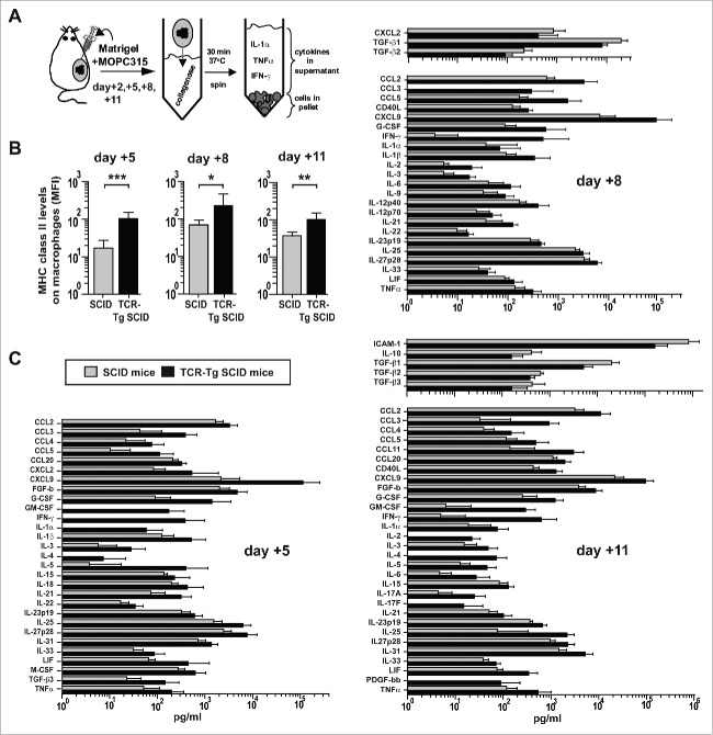
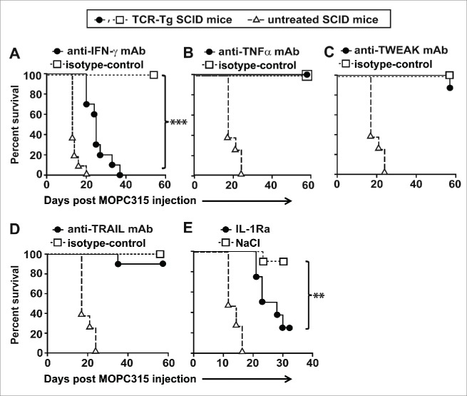
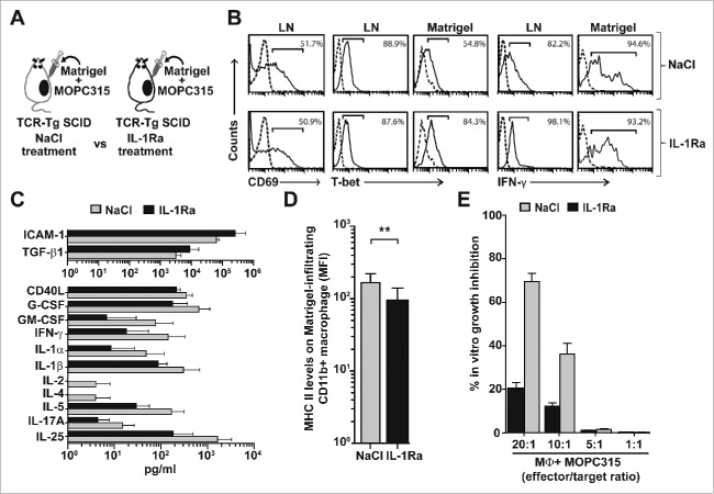
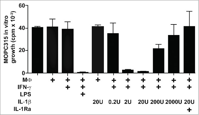
The role of inflammation in cancer is controversial as both tumor-promoting and tumor-suppressive aspects of inflammation have been reported. In particular, it has been shown that pro-inflammatory cytokines, like interleukin-1α (IL-1α), IL-1β, IL-6, and tumor necrosis factor α (TNFα), may either promote or suppress cancer. However, the cellular and molecular basis underlying these opposing outcomes remains enigmatic. Using mouse models for myeloma and lymphoma, we have recently reported that inflammation driven by tumor-specific T helper 1 (Th1) cells conferred protection against B-cell cancer and that interferon-γ (IFN-γ) was essential for this process. Here, we have investigated the contribution of several inflammatory mediators. Myeloma eradication by Th1 cells was not affected by inhibition of TNF-α, TNF-related weak inducer of apoptosis (TWEAK), or TNF-related apoptosis-inducing ligand (TRAIL). In contrast, cancer elimination by tumor-specific Th1 cells was severely impaired by the in vivo neutralization of both IL-1α and IL-1β (collectively named IL-1) with IL-1 receptor antagonist (IL-1Ra). The antitumor functions of tumor-specific Th1 cells and tumor-infiltrating macrophages were both affected by IL-1 neutralization. Secretion of the Th1-derived cytokines IL-2 and IFN-γ at the incipient tumor site was severely reduced by IL-1 blockade. Moreover, IL-1 was shown to synergize with IFN-γ for induction of tumoricidal activity in tumor-infiltrating macrophages. This synergy between IL-1 and IFN-γ may explain how inflammation, when driven by tumor-specific Th1 cells, represses rather than promotes cancer. Collectively, the data reveal a central role of inflammation, and more specifically of the canonical pro-inflammatory cytokine IL-1, in enhancing Th1-mediated immunity against cancer.

Keywords: CD4+ T cells; IFN-γ; IL-1RA; IL-1α; IL-1β; IL-21; IL-27; Th1; and IL33; cancer immunosurveillance; inflammation; macrophages.


Figures

Figure 1.
Figure 1.
Cytokine profile of myeloma eradication mediated by tumor-specific Th1 cells. (A) Experimental design. MOPC315 myeloma cells were mixed with cold liquid Matrigel before s.c. injection into idiotype-specific TCR-Tg SCID or control non-transgenic SCID mice. At body temperature, the Matrigel solution forms a gel plug that contains the myeloma cells, infiltrating immune cells, and locally secreted cytokines. At indicated time points, the Matrigel plugs were excised, dissolved with collagenase, and centrifuged. Cells in the pellet were analyzed by flow cytometry. Extracellular cytokines in the Matrigel were quantified by Luminex technology. (B) MHC class II levels on Matrigel-infiltrating CD11b+ macrophages in TCR-Tg SCID or control SCID mice at indicated time points (mean ±S.D, n = 8−12 mice per group). MFI, mean fluorescence intensity. *p = 0.02, **p = 0.001, *** p < 0.0001 (Mann-Whitney test). (C) The concentration of 46 cytokines in Matrigel supernatant was measured for control SCID (gray bars) or TCR-Tg SCID (black bars) mice (n = 8−12). Only cytokines with significantly higher levels (p < 0.05, Mann-Whitney test) in one group compared with the other are included in the bar graphs (mean ± SD). Data are representative for two independent experiments.
Figure 2.
Figure 2.
Both IFN-γ and IL-1 are required for myeloma elimination by tumor-specific Th1 cells. Idiotype-specific TCR-Tg SCID (n = 6−10 mice per group) or SCID mice (n = 4−6) were injected s.c. with MOPC315 myeloma cells in PBS. Tumor growth was recorded over time. Mice with a tumor diameter ≥10 mm were euthanized. (A–D) TCR-Tg SCID mice were treated i.p. with blocking mAb against (A) IFN-γ, (B) TNFα, (C) TWEAK, or (D) TRAIL or with isotype-matched control mAb. (E) TCR-Tg SCID mice were implanted s.c. with osmotic pumps releasing IL-1Ra or vehicle (NaCl).
Figure 3.
Figure 3.
Roles of IL-1 for myeloma eradication mediated by tumor-specific Th1 cells. (A) Experimental design. Idiotype-specific TCR-Tg SCID mice were injected s.c. with MOC315 in Matrigel and treated s.c. daily with IL-1Ra or vehicle (NaCl). Draining LN and Matrigel plugs were analyzed at day +8. (B) Tumor-specific CD4+ T cells in pooled draining LN and Matrigel plugs from IL-1Ra or NaCltreated mice (n = 8−10 mice per group) were analyzed by flow cytometry. Tumor-specific T cells were gated using the GB113 mAb specific for the transgenic TCR. Levels of surface CD69 or intracellular T-bet and IFN-γ were recorded (solid lines). Dotted lines indicate isotype-matched control mAb. (C) The concentration of 46 cytokines in the extracellular matrix of the Matrigel plugs was quantified for IL-1Ra-treated (black bars) and NaCltreated (gray bars) TCR-Tg SCID mice (n = 11, mean ± SD). Only cytokines which showed significantly (p < 0.05, Mann–Whitney test) higher levels in one group compared with the other are included in the bar graphs. (D) MHC class II levels on Matrigel-infiltrating CD11b+ cells in NaCl or IL-1Ra-treated TCR-Tg SCID mice (n = 8−10, mean ± SD). **p = 0.003 (Mann–Whitney test). (E) Growth inhibition assay. Matrigel-infiltrating CD11b+ macrophages were isolated from IL-1Ra (black bars) or NaCl (gray bars)-treated TCR-Tg SCID mice (n = 10 mice per group), pooled for each group and tested, at various effector to target ratios, for their ability to suppress the in vitro proliferation of MOPC315 cells (mean of quadruplicates ±S.D). MΦ, macrophages.
Figure 4.
Figure 4.
IFN-γ and IL-1β synergize to induce tumoricidal activity in macrophages. SCID mice (n = 12) were injected with MOPC315 in Matrigel. At day +8, Matrigel-infiltrating CD11b+ macrophages were purified using mAb-conjugated magnetic beads, pooled, treated as indicated (with IFN-γ, LPS, various concentrations of IL-1β, and IL-1Ra), and tested for their ability to suppress the in vitro proliferation of MOPC315 (effector to target ratio 10:1). MΦ, macrophages. Data are presented as mean of triplicates ±SD and representative for two independent experiments.

References

    1. Corthay A, Skovseth DK, Lundin KU, Rosjo E, Omholt H, Hofgaard PO, Haraldsen G, Bogen B. Primary antitumor immune response mediated by CD4+ T cells. Immunity 2005; 22:371-83; PMID:15780993; http://dx.doi.org/ 10.1016/j.immuni.2005.02.003 - DOI - PubMed
    1. Galon J, Costes A, Sanchez-Cabo F, Kirilovsky A, Mlecnik B, Lagorce-Pages C, Tosolini M, Camus M, Berger A, Wind P et al.. Type, density, and location of immune cells within human colorectal tumors predict clinical outcome. Science 2006; 313:1960-4; PMID:17008531; http://dx.doi.org/ 10.1126/science.1129139 - DOI - PubMed
    1. Kristensen VN, Vaske CJ, Ursini-Siegel J, Van Loo P, Nordgard SH, Sachidanandam R, Sorlie T, Warnberg F, Haakensen VD, Helland A et al.. Integrated molecular profiles of invasive breast tumors and ductal carcinoma in situ (DCIS) reveal differential vascular and interleukin signaling. Proc Natl Acad Sci U S A 2012; 109:2802-7; PMID:21908711; http://dx.doi.org/ 10.1073/pnas.1108781108 - DOI - PMC - PubMed
    1. Tran E, Turcotte S, Gros A, Robbins PF, Lu YC, Dudley ME, Wunderlich JR, Somerville RP, Hogan K, Hinrichs CS et al.. Cancer immunotherapy based on mutation-specific CD4+ T cells in a patient with epithelial cancer. Science 2014; 344:641-5; PMID:24812403; http://dx.doi.org/ 10.1126/science.1251102 - DOI - PMC - PubMed
    1. Haabeth OA, Tveita AA, Fauskanger M, Schjesvold F, Lorvik KB, Hofgaard PO, Omholt H, Munthe LA, Dembic Z, Corthay A et al.. How Do CD4(+) T Cells Detect and Eliminate Tumor Cells That Either Lack or Express MHC Class II Molecules? Front Immunol 2014; 5:174; PMID:24782871; http://dx.doi.org/ 10.3389/fimmu.2014.00174 - DOI - PMC - PubMed

Publication types