. 2015 Jun 9;5(1):e1057385.
doi: 10.1080/2162402X.2015.1057385. eCollection 2016.

Ibrutinib enhances IL-17 response by modulating the function of bone marrow derived dendritic cells

Affiliations

Ibrutinib enhances IL-17 response by modulating the function of bone marrow derived dendritic cells

Gayathri Natarajan et al. Oncoimmunology. .

Abstract

Ibrutinib (PCI-32765) is an irreversible dual Btk/Itk inhibitor shown to be effective in treating several B cell malignancies. However, limited studies have been conducted to study the effect of this drug on myeloid cell function. Hence, we studied the effect of ibrutinib treatment on TLR-4 mediated activation of bone marrow derived dendritic cell culture (DCs). Upon ibrutinib treatment, LPS-treated DCs displayed lower synthesis of TNF-α and nitric oxide (NO) and higher induction of IL-6, TGF-β, IL-10 and IL-18. While ibrutinib dampened MHC-II and CD86 expression on DCs, CD80 expression was upregulated. Further, ibrutinib-treated DCs promoted T cell proliferation and enhanced IL-17 production upon co-culture with nylon wool enriched T cells. Taken together, our results indicate that ibrutinib modulates TLR-4 mediated DC activation to promote an IL-17 response. We describe a novel mode of action for ibrutinib on DCs which should be explored to treat other forms of cancer besides B cell malignancies.

Keywords: Btk; IL-17; T cells; TLR-4; dendritic cells; ibrutinib.


Figures

Figure 1.
Figure 1.
Ibrutinib dampens TNF-α and nitric oxide production in dendritic cells upon LPS stimulation. (A) TNF-α, (B) nitric oxide (NO) and (C) IL-12 production in control- and ibrutinib-treated DCs stimulated with LPS. DCs were treated with control (DMSO) or ibrutinib (1 µM or 10 µM), washed twice and treated with LPS (1 µg/mL). After 4, 12 and 24 h of LPS treatment, cytokine production was determined in the culture supernatants by ELISA. At these time points, NO levels were determined in the culture supernatants by measuring nitrite concentrations using Griess assay. The data are presented as mean + SEM of triplicate sample values from 2 independent experiments. *p < 0.05, **p < 0.001, ***p < 0.0001.
Figure 2.
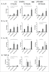
Figure 2.
Ibrutinib enhances the induction of IL-6, IL-10, IL-18 and TGF-β in dendritic cells upon LPS stimulation. (A) IL-6, (B) IL-10, (C) IL-18 and (D) TGF-β mRNA induction in control and ibrutinib-treated DCs upon LPS stimulation. DCs were treated with control (DMSO) or ibrutinib (1 µM or 10 µM), washed twice and treated with LPS (1 µg/mL). After 4, 12 and 24 h of LPS treatment, cells were treated with TRIzol Reagent, RNA was isolated from cells and mRNA levels of respective cytokines were determined by real-time qPCR. The data are presented as mean + SEM of duplicates obtained by pooling 3 samples in 2 independent experiments. *p < 0.05.
Figure 3.
Figure 3.
Ibrutinib differentially regulates the surface expression of MHC-II and co-stimulatory molecules in LPS-treated dendritic cells. Histogram plots show expressions of (A) MHC-II, (B) CD86 and (C) CD80 in LPS/control and LPS/ibrutinib-treated DC cultures. Numbers represent mean percentage of cells + SEM of the respective surface molecule on DCs. The data presented are representative plots of 3 independent experiments. Mean fluorescence intensities (MFIs) of (D) MHC-II, (E) CD86 and (F) CD80 expression in LPS/control and LPS/ibrutinib-treated DC cultures. The data are presented as mean + SEM of representative MFI values of 3 independent experiments. DCs were treated with control (DMSO) or ibrutinib (1 µM), washed twice and treated with LPS (1 µg/mL). After 24 h of LPS treatment, cells were blocked, stained with conjugated antibodies for the respective surface molecules and expressions of the surface molecules were determined by flow cytometry. Analyses were conducted by gating on CD11c+ DCs. *p < 0.05, **p < 0.001, ***p < 0.0001.
Figure 4.
Figure 4.
Ibrutinib treated DCs promote IL-17 response upon culture with T cells. (A) Analysis of T cell proliferation upon co-culture with control-, ibrutinib-, LPS/control- or LPS/ibrutinib-treated DCs. DCs were treated with control (DMSO) or ibrutinib (1 µM), washed twice, pulsed with OVA (10 µg/mL) for 2 h and treated with LPS (1 µg/mL) for 22 h. After OVA/LPS stimulation, DCs were cultured in 1:4 ratio with CFSE-stained T cells enriched from spleens of OT-II mice for 5 d. At day 5, cells from co-culture were blocked, stained with anti-CD4 antibody and T cell proliferation was measured by flow cytometry. Analyses were conducted by gating on CD4+ population. The data are presented as mean + SEM of duplicates and are representative of 2 independent experiments. (B) Production of T cell cytokines IL-17, (C) IFNγ and (D) IL-13 in co-culture experiments performed as mentioned in A. At Day 5 of co-culture, cell culture supernatants were collected and the respective cytokines were measured by ELISA. The data are presented as mean + SEM of duplicates and are representative of 2 independent experiments. *p < 0.05, **p < 0.001.

References

    1. Honigberg LA, Smith AM, Sirisawad M, Verner E, Loury D, Chang B, Li S, Pan Z, Thamm DH, Miller RA et al.. The Bruton tyrosine kinase inhibitor PCI-32765 blocks B-cell activation and is efficacious in models of autoimmune disease and B-cell malignancy. Proc Natl Acad Sci U S A 2010; 107:13075-80; PMID:20615965; http://dx.doi.org/10.1073/pnas.1004594107 - DOI - PMC - PubMed
    1. Dubovsky JA, Beckwith KA, Natarajan G, Woyach J, Jaglowski S, Zhong Y, Hessler JD, Liu T-M, Chang BY, Larkin KM et al.. Ibrutinib is an irreversible molecular inhibitor of ITK driving a Th1-selective pressure in T lymphocytes. Blood 2013; 122:2539-49; PMID:23886836; http://dx.doi.org/10.1182/blood-2013-06-507947 - DOI - PMC - PubMed
    1. Akinleye A, Chen Y, Mukhi N, Song Y, Liu D. Ibrutinib and novel BTK inhibitors in clinical development. J Hematol Oncol 2013; 6:59; PMID:23958373; http://dx.doi.org/10.1186/1756-8722-6-59 - DOI - PMC - PubMed
    1. Maddocks K, Blum KA. Ibrutinib in B-cell Lymphomas. Curr Treat Options Oncol 2014; 15:226-37; PMID:24481980; http://dx.doi.org/10.1007/s11864-014-0274-8 - DOI - PubMed
    1. Burger JA, Buggy JJ. Bruton tyrosine kinase inhibitor ibrutinib (PCI-32765). Leuk Lymphoma 2013; 54:2385-91; PMID:23425038; http://dx.doi.org/10.3109/10428194.2013.777837 - DOI - PubMed

Publication types