

Review
doi: 10.1016/j.molcel.2016.01.031.
The Emerging Network of Mitochondria-Organelle Contacts
Affiliations
- PMID: 26942669
- PMCID: PMC5554544
- DOI: 10.1016/j.molcel.2016.01.031
Item in Clipboard
Review
The Emerging Network of Mitochondria-Organelle Contacts
Mol Cell.
.
Abstract
Membrane contact sites between mitochondria and other organelles are important for lipid and ion exchange, membrane dynamics, and signaling. Recent advances are revealing their molecular features and how different types of mitochondria contacts are coordinated with each other for cell function.
Copyright © 2016 Elsevier Inc. All rights reserved.
Figures
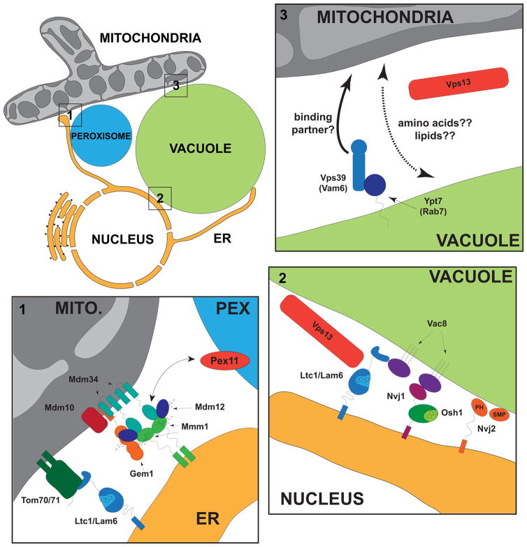
Current understanding of proteins localized to contact sites between mitochondria (gray), vacuoles (green), ER (yellow) and peroxisomes (blue) in budding yeast. Detailed depictions of ER-mitochondria-peroxisome, nucleus –vacuole and mitochondria-vacuole contacts are shown in enlargements 1–3, respectively. ERMES and Ltc1 localize independently to ER-mitochondria contacts, shown in the bottom left enlargement. Ltc1 interacts with Tom70 and Tom71 via its GRAM domain (light blue) and is anchored into the ER via its predicted transmembrane domain. In ERMES, Mmm1 (green) homodimers are flanked by single Mdm12 SMP domains (purple) and this tetramer interacts with Mdm34 (turquoise) to potentially transport PC from the ER to mitochondria. The roles and interactions of the core ERMES and peripheral ERMES subunits, Mdm10 and Gem1, respectively, are not known. Peroxisomes are adjacent to ER-mitochondria contacts, possibly thorough an interaction with Mdm34. Enlarged in the top left are vCLAMPs and shown schematically are Vps39 and Ypt7, which are required for their formation, and Vps13. Both Vps39 and Vps13 are enriched in vCLAMP regions and Vps13 may reinforce vCLAMP formation. Whether specific mitochondrial components are required for vCLAMP formation or are enriched at these regions as part of a vCLAMP tether complex is not known. Enlarged in the lower right corner are types of ER-vacuolar contacts. Nuclear-vacuolar junctions (NVJs) are sites at which starvation-induced piecemeal microautophagy of the nuclear envelope occurs and are formed by a physical interaction between Nvj1 and Vac8. NVJs also contain Osh1, a sterol transport protein and the PH and SMP-domain containing protein, Nvj2, which binds to membranes, but its lipid ligands are not well established. Vac8 is also required to localize Ltc1/Lam6 to these sites and to non-NVJ ER-vacuole contacts required for stress induced vacuolar membrane domain formation. The conserved protein, Vps13 is localized to ER-vacuolar contacts in a regulated manner under respiratory growth conditions.
References
-
- Cohen Y, Klug YA, Dimitrov L, Erez Z, Chuartzman SG, Elinger D, Yofe I, Soliman K, Gartner J, Thoms S, et al. Peroxisomes are juxtaposed to strategic sites on mitochondria. Molecular bioSystems. 2014;10:1742–1748. - PubMed
-
- de Brito OM, Scorrano L. Mitofusin 2 tethers endoplasmic reticulum to mitochondria. Nature. 2008;456:605–610. - PubMed
-
- Elbaz-Alon Y, Rosenfeld-Gur E, Shinder V, Futerman AH, Geiger T, Schuldiner M. A dynamic interface between vacuoles and mitochondria in yeast. Developmental cell. 2014;30:95–102. - PubMed
Publication types
MeSH terms
Grants and funding
LinkOut - more resources
Full Text Sources
Other Literature Sources
