Review
. 2016 Mar 3;61(5):654-666.
doi: 10.1016/j.molcel.2016.01.028.

The Mitochondrial Basis of Aging

Affiliations
Review

The Mitochondrial Basis of Aging

Nuo Sun et al. Mol Cell. .

Abstract

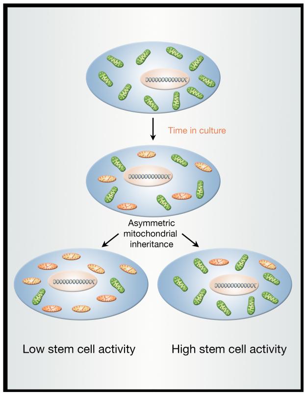
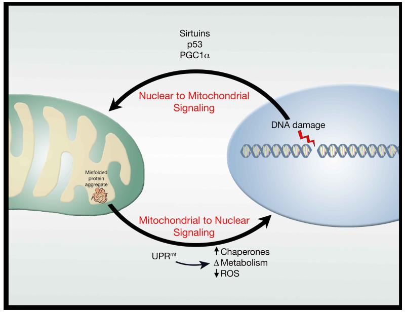
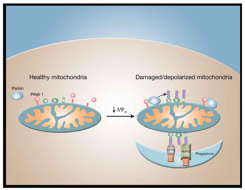
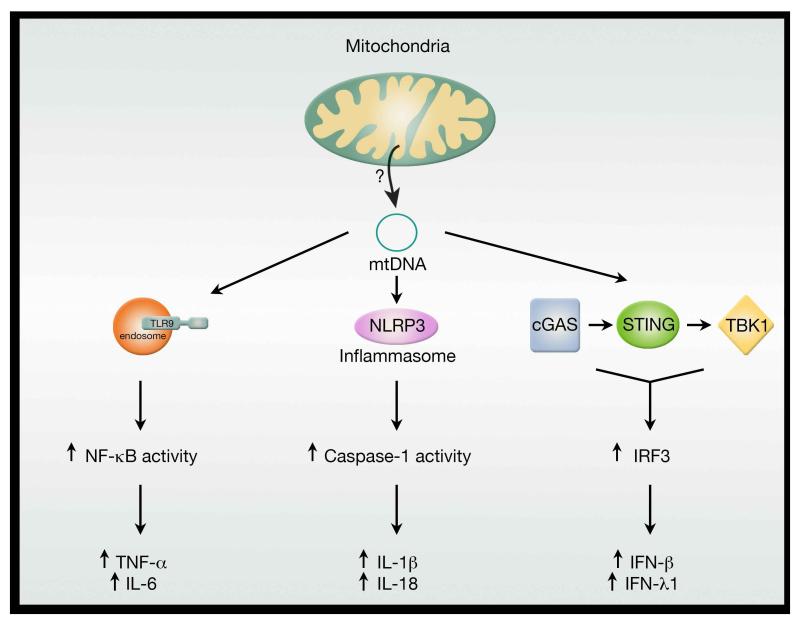
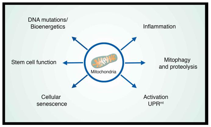
A decline in mitochondrial quality and activity has been associated with normal aging and correlated with the development of a wide range of age-related diseases. Here, we review the evidence that a decline in mitochondria function contributes to aging. In particular, we discuss how mitochondria contribute to specific aspects of the aging process, including cellular senescence, chronic inflammation, and the age-dependent decline in stem cell activity. Signaling pathways regulating the mitochondrial unfolded protein response and mitophagy are also reviewed, with particular emphasis placed on how these pathways might, in turn, regulate longevity. Taken together, these observations suggest that mitochondria influence or regulate a number of key aspects of aging and suggest that strategies directed at improving mitochondrial quality and function might have far-reaching beneficial effects.


Figures

Figure 1
Figure 1
Stem cells exhibit asymmetric mitochondrial inheritance. Analysis of stem-like cells within immortalized, transformed epithelial cultures revealed that young mitochondria (shown in green) and old mitochondria (depicted in orange) are not symmetrically distributed after the stem-like cell divides. Moreover, the daughter cell inheriting the younger mitochondria, also exhibits higher stem-like activity. The molecular basis for this asymmetric mitochondrial distribution is not clear, nor is it known whether similar mechanisms exist in vivo.
Figure 2
Figure 2
Bidirectional signaling between the nucleus and mitochondria. Communication exists between the nucleus and the mitochondria with evidence that nuclear stresses, such as DNA damage, trigger a mitochondrial response. Similarly, mitochondrial stresses, such as protein aggregates, stimulate a retrograde response to the nucleus. Both directions of this signaling paradigm appear intimately linked to longevity.
Figure 3
Figure 3
Parkin-dependent mitophagy. In healthy mitochondria, the PINK1 kinase is constitutively degraded. A fall in mitochondrial membrane potential (Δψm) stabilizes PINK1 facilitating the recruitment of cytosolic Parkin to the mitochondrial outer membrane. Activation of Parkin results in the ubiquitinization (purple balls) of multiple outer mitochondrial membrane proteins (shown in green). Once ubiquinated, these proteins are recognized by specific mitophagy receptors such as optineurin (OPTN) and NDP52, which along with LC3, directs the phagophore to surround the damaged mitochondria allowing for its ultimate delivery to the lysosome for degradation via mitophagy.
Figure 4
Figure 4
Mitochondria as regulators of the innate immune response. Release of mitochondrial DNA appears to trigger at least three distinct pathways linked to inflammation. The precise mechanism by which free mitochondrial DNA enters the cytosol to engage with various intracellular DNA sensors is currently unclear. Nonetheless, age-dependent breakdown of the mitochondrial membrane might allow escape of mtDNA and thereby help fuel the chronic, sterile inflammation associated with aging.
Figure 5
Figure 5
Mitochondria as regulators of organismal aging. The contribution of mitochondria to the aging process occurs through multiple distinct pathways. Although depicted as separate pathways, clear intersections occur as is evident between the connection between activation of the UPRmt and the induction of the inflammatory response (see text for details).

Similar articles

Cited by

References

    1. Ahlqvist KJ, Hamalainen RH, Yatsuga S, Uutela M, Terzioglu M, Gotz A, Forsstrom S, Salven P, Angers-Loustau A, Kopra OH, et al. Somatic progenitor cell vulnerability to mitochondrial DNA mutagenesis underlies progeroid phenotypes in Polg mutator mice. Cell Metab. 2012;15:100–109. - PubMed
    1. Arany Z, He H, Lin J, Hoyer K, Handschin C, Toka O, Ahmad F, Matsui T, Chin S, Wu PH, et al. Transcriptional coactivator PGC-1 alpha controls the energy state and contractile function of cardiac muscle. Cell Metab. 2005;1:259–271. - PubMed
    1. Argiles JM, Busquets S, Stemmler B, Lopez-Soriano FJ. Cachexia and sarcopenia: mechanisms and potential targets for intervention. Curr Opin Pharmacol. 2015;22:100–106. - PubMed
    1. Austin S, St-Pierre J. PGC1alpha and mitochondrial metabolism--emerging concepts and relevance in ageing and neurodegenerative disorders. J Cell Sci. 2012;125:4963–4971. - PubMed
    1. Bennett CF, Vander Wende H, Simko M, Klum S, Barfield S, Choi H, Pineda VV, Kaeberlein M. Activation of the mitochondrial unfolded protein response does not predict longevity in Caenorhabditis elegans. Nat Commun. 2014;5:3483. - PMC - PubMed

Publication types