. 2016 Mar 3;61(5):720-733.
doi: 10.1016/j.molcel.2016.02.007.

TRIM21 Ubiquitylates SQSTM1/p62 and Suppresses Protein Sequestration to Regulate Redox Homeostasis

Affiliations

TRIM21 Ubiquitylates SQSTM1/p62 and Suppresses Protein Sequestration to Regulate Redox Homeostasis

Ji-An Pan et al. Mol Cell. .

Erratum in

Abstract

TRIM21 is a RING finger domain-containing ubiquitin E3 ligase whose expression is elevated in autoimmune disease. While TRIM21 plays an important role in immune activation during pathogen infection, little is known about its inherent cellular function. Here we show that TRIM21 plays an essential role in redox regulation by directly interacting with SQSTM1/p62 and ubiquitylating p62 at lysine 7 (K7) via K63-linkage. As p62 oligomerizes and sequesters client proteins in inclusions, the TRIM21-mediated p62 ubiquitylation abrogates p62 oligomerization and sequestration of proteins including Keap1, a negative regulator of antioxidant response. TRIM21-deficient cells display an enhanced antioxidant response and reduced cell death in response to oxidative stress. Genetic ablation of TRIM21 in mice confers protection from oxidative damages caused by arsenic-induced liver insult and pressure overload heart injury. Therefore, TRIM21 plays an essential role in p62-regulated redox homeostasis and may be a viable target for treating pathological conditions resulting from oxidative damage.


Figures

Figure 1
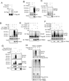
Figure 1. p62 is ubiquitylated at K7 via the K63-linkage
(A) Baby mouse kidney (BMK) cells were transfected with His-Ub for 24 h, then treated with MG132 (0.25 µM) and subjected to His pull-down (Ni-NTA) and immunoblotting (IB). (B) Flag- and His-doubly tagged p62wt, K7R, and K13R were transfected into HEK293T cells for 36 h. Cells were subjected to His-pull down and IB. (C) Flag and His-doubly tagged p62wt and K7R were co-transfected with HA-Ub wt, K48-only, and K63-only mutants in HEK293T cells. Cells were subjected to His pull-down and IB. (D) Indicated p62 mutants were co-expressed in HEK293T cells. Cells were subjected to His-pull down and IB. (E) Flag-tagged p62wt or indicated mutants were expressed in HEK293T cells. HA-Ub was expressed in another set of HEK293T cells. 24 h post transfection, p62 or mutant-expressing cell lysates were incubated with the same amount of the HA-Ub expressing cell lysates for 3 h. The mixtures were subjected to IP with FLAG antibody then IB with HA antibody. (F) p62−/− MEFs stably expressing GFP-LC3 were reconstituted with p62wt and p62K7R. Cells were treated with MG132 (0.25 µM) for 12 h, then observed under deconvolution microscope. The percentage of cells with GFP-LC3 aggregates was quantified by counting over 50 cells in each sample. Data shown are the mean plus S.D. of three countings. ***p<0.01.
Figure 2
Figure 2. p62 is ubiquitylated by TRIM21
(A) Flag-p62 was expressed in HEK293T cells, and subjected to IP using the Flag antibody and eluted using Flag peptides. The eluates were incubated with in vitro ubiquitination assay system containing E1, E2, and ubiquitin, then subjected to IB. (B and C) His-Ub was transfected with vector and HA-TRIM21 (B) or HA-RNF138 (C) into HEK293T cells. 24 h later, cells were subjected to His pull-down followed by IB. (D) His-Ub was co-transfected with HA-TRIM21wt or HA-TRIM21-LD mutant in HEK293T cells. Cells were subjected to His pull-down and IB. (E) Purified Flag-p62 was incubated with IgG control, purified HA-TRIM21wt or LD mutant in the ubiquitination assaying mix, and probed for indicated proteins. (F) His-Ub (wt, K48R, and K63R) mutants were expressed with or without HA-TRIM21 in HEK293T cells. Cells were subjected to His pull-down and IB. (G) Flag- and His-doubly tagged p62wt or K7R mutant were co-expressed with vector or TRIM21 in HEK293T cells. Cells were subjected to His-pull down and IB. (H) MD-MBA-231 with stable expression of Flag-p62-His were infected with lentiviral non-targeting control (NTC) shRNA or shTRIM21. Cells were treated with MG132, then subjected to His pull-down and IB.
Figure 3
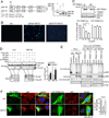
Figure 3. TRIM21 inhibits p62 dimerization and its sequestration function
(A) Schematic for the strategy constructing p62 fused with Venus N-terminus (p62-VN) or C-terminus (p62-VC). p62-VN and p62-VC were expressed individually or simultaneously in HEK293T cells. The expression was determined by IB. (B) Wild-type or K7R p62-VN/p62-VC were expressed in HEK293T cells. Representative fluorescence microscopic images are shown. (C) p62-VN or p62-VC were expressed with estrogen receptor (ER)-fused TRIM21wt or LD mutant in HEK293T cells. 16 h later, cells were treated with 4-OHT for indicated times to induce TRIM21 expression (top panel). Venus fluorescence was determined by flow cytometry and quantified (bottom panel). (D) HeLa cells stably expressing both Flag- and HA-p62 were infected with lentivirus shNTC and shTRIM21. Cells were used for Flag IP, and IB for HA. The amount of HA-p62 was quantified by ImageJ and is shown in the right panel. *p<0.05. (E) HeLa with stable shTRIM21 were reconstituted with shRNA-resistant TRIM21wt or TRIM21 LD. Cells treated with MG132, then crosslinked with DSP, and lysed in IP lysis buffer with 1% SDS. The cells lysates mixed with loading buffer containing or not containing β-mercaptoethanol were subjected to IB. (F) TRIM21−/− MEFs reconstituted with TRIM21wt or LD were treated with MG132 (0.25 µM) for 12 h. Cells were used for IF using anti-TRIM21 (green) and anti-p62 (red) antibodies, and observed under deconvolution microscope. Cells with aggregates were quantified by blind counting. Shown on the right is the mean plus S.D. of three countings. *p<0.05, **p<0.01. Cells with high expression of TRIM21 wt or LD are marked with an asterisk.
Figure 4
Figure 4. TRIM21 interacts with p62 via the PRYSPRY domain
(A–C) Flag-tagged TRIM21 and HA-tagged p62wt or their truncation mutants were expressed in indicated combinations in HEK293T cells for 24 h. Cells were subjected to Flag IP and IB. p62 lost interaction with TRIM21 Δ213–117 and Δ117–200 (A). TRIM21 lost interaction with p62 Δ3–61, Δ117–162, and Δ163–200 (B). p62 lost interaction with TRIM21 Δ252–476 (C). (D) Flag-tagged TRIM21wt or W381/383A mutant and HA-tagged p62 were expressed in HEK293T cells for 24 h. Cells were subjected to Flag IP and IB. (E) HA-tagged TRIM21wt or W381/383A mutant and His-Ub were expressed in HEK293T cells. Cells were subjected to His pull-down and IB. (F) TRIM21−/− MEFs reconstituted with vector, TRIM21wt, and TRIM21 W381/383A mutant were treated with DSP and lysed in IP lysis buffer with 1% SDS. Lysates were mixed with loading buffer with/without β-mercaptoethanol and subjected to IB. (G) HA-tagged p62 and Flag-tagged TRIM21 were expressed in HEK293T cells for 24 h. Cells were treated with MG132 (0.5 µM) for 16 h, rapamycin (50 nM) for 16 h, tunicamycin (0.5 µg/ml) for 16 h, or cultured in serum-free medium for 6 h. Cells were subjected to Flag IP and IB. (H) A vector control or Flag tag was introduced into MEFs to fuse with the endogenous TRIM21 by CRISPR. Cells were treated with MG132 (0.25 µM) for 12 h, then subjected to Flag IP and IB.
Figure 5
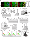
Figure 5. Trim21 negatively regulates p62-mediated Keap1 sequestration and antioxidant response
(A) p62−/− MEFs reconstituted with vector, Flag-p62wt, or Flag-p62K7R mutant were untreated or treated with sodium arsenate (As(III), 10 µM) for 6 h. Cells were subjected to IF with Flag and Keap1 antibodies, and observed under deconvolution microscope. Cells with Keap1 aggregates were counted blindly. Data shown are the averages plus S.D. of at least three countings with over 200 cells. **p<0.01, ***p<0.001. (B) p62−/− MEFs reconstituted with vector, Flag-p62wt, and Flag-p62K7R were treated with As(III) (10 µM) for 2.5 h. Cells were lysed in IP lysis buffer with 1% Triton X-100. The insoluble fraction was lysed in IP lysis buffer with 1% SDS. 30 µg soluble proteins and corresponding volume of insoluble proteins were used for IB. (C) p62−/− MEFs reconstituted with vector, Flag-p62wt, and Flag-p62K7R were left untreated or treated with As(III) (10 µM) for 4 h. Cells were treated with cycloheximide (CHX) for indicated times. Cells were lysed in RIPA (1% SDS) and probed for Nrf2 and tubulin. (D) p62−/− MEFs reconstituted with vector, Flag-p62wt, and Flag-p62K7R were transfected with NQO1-ARE-luc and pRL-TK (Renilla Luciferase Control Reporter Vector, Promega) plasmids. 16 h post transfection, cells were treated with As(III) and assayed for luciferase activity. (E) MEFs without/with CRISPR Flag fused into TRIM21 gene were treated with As(III) (10 µM) for 4 h, and subjected to Flag IP and IB. (F) TRIM21−/− MEFs reconstituted with vector, TRIM21wt, and TRIM21W381/383A mutant were treated with the crosslinking agent DSP and lyse in IP lysis buffer with 1% SDS. The lysates were mixed with loading buffer with/without β-mercaptoethanol and subjected to SDS-PAGE and IB. (G) TRIM21−/− MEFs reconstituted with vector and HA-TRIM21wt were treated with As(III). Cells were lysed in IP lysis buffer containing 1% Triton X-100. The insoluble fraction was dissolved in RIPA buffer containing 1% SDS. Both the Triton X-100 soluble and insoluble fractions were subjected to IB. (H) TRIM21−/− MEFs reconstituted with vector and HA-TRIM21wt were treated with As(III) (10 µM). Cell were fractionated to obtain the cytosol and nuclear fractions, and probed for indicated proteins. (I) TRIM21−/− MEFs reconstituted with vector, HA-TRIM21wt, or HATRIM21W381/ 383A mutants were treated with As(III) (10 µM) for 4 h. Cells were lysed in RIPA buffer containing 1% SDS and probed for indicated proteins. (J) TRIM21−/− MEFs reconstituted with vector, HA-TRIM21wt, HA-TRIM21-LD, or HA-TRIM21W381/383A were transfected with NQO1-ARE-luc and renilla luciferase pRL-TK constructs. 16 h later, cells were treated with As(III) (10 µM), and assayed for luciferase activity. (K) TRIM21−/− MEFs reconstituted with vector, HA-TRIM21wt, HA-TRIM21-LD, or HA-TRIM21W381/383A mutants were treated with As(III) (10 µM) for 4 h. Cells were stained with H2DCFDA and analyzed by flow cytometry. Quantification of relative H2DCFDA intensity (Geo mean plus S.D. of four repeats) is shown on the right. ***p<0.001, ****p<0.0001.
Figure 6
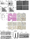
Figure 6. Ablation of TRIM21 led to the protection from arsenic toxicity
(A and B) TRIM21+/+ and −/− MEFs were treated with As(III) (10 µM) for 12 h (A), or for the indicated times (B). Cells were collected and the lysates were subjected to IB. (C and D) MEFs were treated with As(III) (10 µM). (C) Cells were observed under phase-contrast microscope 16 h after the treatment. Representative micrographs are shown. (D) Cell viability was determined by Trypan Blue. Data shown are the mean plus S.D. of three countings. **p<0.01. (E) TRIM21−/− MEFs reconstituted with vector, HA-TRIM21wt, or HA-TRIM21W381/383A mutant were treated with As(III) (10 µM). Cell viability was determined by Trypan Blue. Shown are the mean plus S.D. of three countings. **p<0.01. (F–I) Nine pairs of litter mates of TRIM21+/+ and −/− mice were gavaged with 30 mg/kg As2O3. (F) Body weight was measured before and after 24 h treatment, and loss of body weight was calculated and plotted. *p<0.05. (G) Livers from water or As2O3-treated mice were collected after 24 h. Tissues were processed for H&E staining and IHC. Representative images are shown. (H) TRIM21+/+ and TRIM21−/− mice were treated with As2O3 for 24 h. Liver tissues were homogenized and separated into detergent (1% Triton X-100)-soluble “S” and insoluble “I” fractions, and subjected to IB. The ratio of insoluble/soluble band intensities was normalized to the corresponding tubulin band, and is shown on the right. *p<0.05, **p<0.01. (I) Liver tissue was lysed in RIPA buffer containing 1% SDS and subjected to IB.
Figure 7
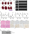
Figure 7. Ablation of TRIM21 leads to protection against heart injury induced by sTAC
Sham or sTAC was performed on wild-type or TRIM21−/− mice. 4 d later, animals were subjected to echocardiography, then the hearts were isolated for further analyses. (A) Hearts from each group were photographed. (B) Representative B-mode (left ventricle labeled) and M-mode echocardiographic images are shown. (C) M-mode images from 9 cardiac cycles for each animal were used to calculate ventricular measurements: LVDV (left ventricular diastolic volume), LVSV (left ventricular systolic volume), fractional shortening, FS (fractional shortening) and EF (ejection fraction). Individual data points and the mean are shown. n.s.: nonsignificant; *p<0.05; **p<0.01; ***p<0.001; ****p<0.0001. (D) Hearts from sham or sTAC-treated mice were collected and examined with H&E and IHC staining. (E) Heart tissue was lysed in RIPA buffer containing 1% SDS and subjected to IB.

References

    1. Barchowsky A, Dudek EJ, Treadwell MD, Wetterhahn KE. Arsenic induces oxidant stress and NF-kappa B activation in cultured aortic endothelial cells. Free Radic Biol Med. 1996;21:783–790. - PubMed
    1. Belch JJ, Bridges AB, Scott N, Chopra M. Oxygen free radicals and congestive heart failure. British heart journal. 1991;65:245–248. - PMC - PubMed
    1. Ben-Chetrit E, Chan EK, Sullivan KF, Tan EM. A 52-kD protein is a novel component of the SS-A/Ro antigenic particle. J Exp Med. 1988;167:1560–1571. - PMC - PubMed
    1. Ben-Chetrit E, Fox RI, Tan EM. Dissociation of immune responses to the SS-A (Ro) 52-kd and 60-kd polypeptides in systemic lupus erythematosus and Sjogren's syndrome. Arthritis and rheumatism. 1990;33:349–355. - PubMed
    1. Bernstam L, Nriagu J. Molecular aspects of arsenic stress. Journal of toxicology and environmental health Part B, Critical reviews. 2000;3:293–322. - PubMed

Publication types

MeSH terms