. 2016 Mar 3;61(5):760-773.
doi: 10.1016/j.molcel.2016.02.013.

A Specialized Mechanism of Translation Mediated by FXR1a-Associated MicroRNP in Cellular Quiescence

Affiliations

A Specialized Mechanism of Translation Mediated by FXR1a-Associated MicroRNP in Cellular Quiescence

Syed I A Bukhari et al. Mol Cell. .

Abstract

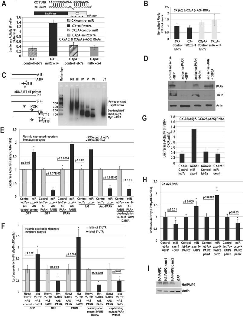
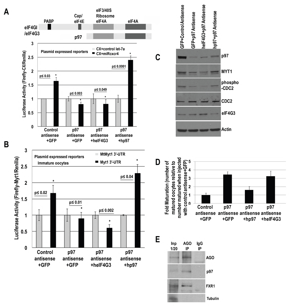
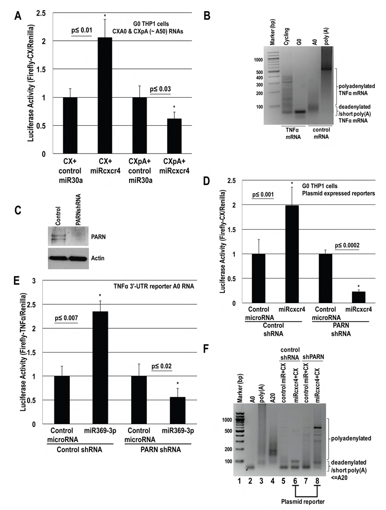
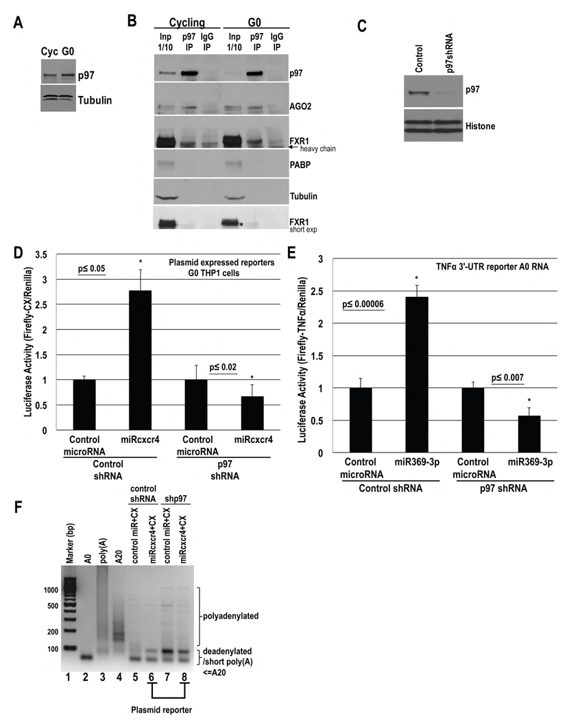
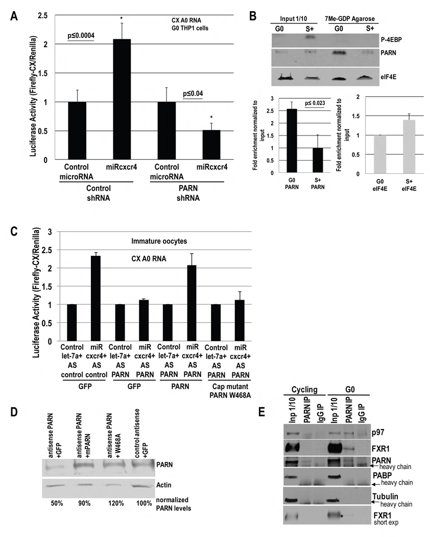
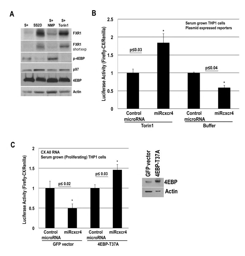
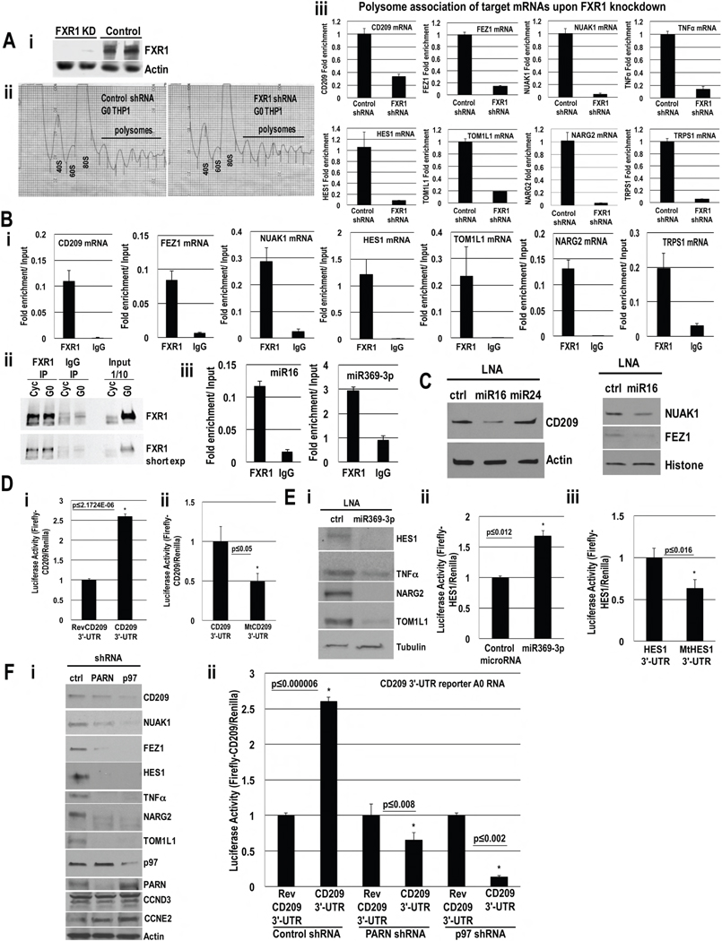
MicroRNAs predominantly decrease gene expression; however, specific mRNAs are translationally upregulated in quiescent (G0) mammalian cells and immature Xenopus laevis oocytes by an FXR1a-associated microRNA-protein complex (microRNP) that lacks the microRNP repressor, GW182. Their mechanism in these conditions of decreased mTOR signaling, and therefore reduced canonical (cap-and-poly(A)-tail-mediated) translation, remains undiscovered. Our data reveal that mTOR inhibition in human THP1 cells enables microRNA-mediated activation. Activation requires shortened/no poly(A)-tail targets; polyadenylated mRNAs are partially activated upon PAIP2 overexpression, which interferes with poly(A)-bound PABP, precluding PABP-enhanced microRNA-mediated inhibition and canonical translation. Consistently, inhibition of PARN deadenylase prevents activation. P97/DAP5, a homolog of canonical translation factor, eIF4G, which lacks PABP- and cap binding complex-interacting domains, is required for activation, and thereby for the oocyte immature state. P97 interacts with 3' UTR-binding FXR1a-associated microRNPs and with PARN, which binds mRNA 5' caps, forming a specialized complex to translate recruited mRNAs in these altered canonical translation conditions.


Conflict of interest statement

The authors declare no conflict of interest.

Figures

Fig. 1
Fig. 1. MicroRNA-mediated activation requires poly(A) shortened mRNAs and PARN to avoid PABP in immature oocytes
A. Luciferase assay of in vitro transcribed capped CX mRNAs (CX=Firefly Luciferase reporter with 4 target sites for artificial microRNA, miRcxcr4, in its 3′-UTR), without/with a poly(A) tail (CX(A0)=CX A0=no poly(A) tail, CXpA=~A50, Fig. S1A), which were injected into stage IV oocyte nuclei with Renilla mRNA and miRcxcr4 or control let-7a. B. qRT-PCR of RNAs in A. C. RACE-PAT assay of endogenous target Myt1 mRNA in immature oocyte stages; oligo dT/RNase H treated RNA=deadenylated RNA control. D.–E. Oocytes were injected with: control antisense (AS control)+GFP, PARN antisense (AS PARN)+GFP, PARN antisense+ PARN (mouse PARN cDNA insensitive to antisense against Xenopus PARN) and PARN antisense+PARN D285A deadenylase inactive mutant, followed by: D. Western blot of PARN and endogenous target MYT1, and E. these oocytes, as well as oocytes injected with control IgG or anti-PARN antibody, were injected with CX and Renilla plasmids and miRcxcr4 or let-7a, followed by Luciferase analysis. PAT assays of reporters (Fig. S1Bi–ii) and endogenous Myt1 mRNA (Fig. S1Biii–iv) reveal altered poly(A) tails; RNA levels do not correlate with translation/poly(A) changes (Fig. S1C–D). F. Oocytes were injected with antisense and rescue plasmids as noted and then with Firefly Luciferase plasmids with the 3′-UTR of endogenous miR16 target, Myt1, or with the 3′-UTR miR16 target site mutated (MtMyt1 3′-UTR) and Renilla, followed by Luciferase assay. G. CXA5 and CXA25 mRNAs (CX mRNAs with A5 and A25, Fig. S1A), Renilla mRNA and miRcxcr4 or let-7a were injected into oocytes, followed by Luciferase assay to test activation with poly(A) tails less than or greater than a PABP binding site (~20 As). H. Oocytes were injected with GFP, HA-tagged PAIP2, PAIP2-pam 1or PAIP2-pam 2 (pam 1 deleted) cDNAs and with CXA25 mRNA, Renilla and miRcxcr4 or let-7a, followed by Luciferase assay to test if PAIP2 enables activation with CXA25. I. Western blot of samples in H. Actin=loading control. Graphs=average of ≥ 3 experiments; error bars=standard error of mean (SEM) or standard deviation (A, G); p≤0.05 or indicated. See Fig. S1.
Fig. 2
Fig. 2. P97 mediates activation and is involved in the immature oocyte state
P97, eIF4GI, eIF4G3 (eIF4G3/eIF4GII, distinct from eIF4G2/DAP5/p97) domains. Oocytes were injected with control or p97 antisense and GFP, human p97 or eIF4G3 rescue cDNA plasmids for 6–7 hr and then with: A. CX and Renilla plasmids, and miRcxcr4 or control let-7a or B. Myt1 3′-UTR or mtMyt1 3′-UTR Firefly Luciferase reporters and Renilla plasmids. C. Western blot of p97, MYT1, and immaturity marker, phospho-CDC2 (MYT1 phosphorylates CDC2), upon p97 depletion and rescue in A. 57% and 61% decrease in MYT1, and 65% and 64% decrease in phospho-CDC2, were observed with p97 depletion and upon lack of rescue with eIF4G3; 94% rescue of MYT1 and 84% rescue of phospho-CDC2 was observed with human p97 compared to control antisense. Actin=loading control. RNA levels do not change significantly (Fig. S2A–B). D. Maturation upon p97 depletion and rescue (samples in A) by scoring for Germinal Vesicle Breakdown (GVBD). Y axis=fold maturation, which is the number of matured oocytes in samples injected with: p97 antisense+GFP, p97 antisense+human p97 (hp97), or p97 antisense+human eIF4G3 (heIF4G3), compared to number of oocytes matured when injected with control antisense+GFP (set to 1). E. Immunoprecipitation (IP) with AGO antibody and control IgG from RNAse treated oocyte extracts; Western blot for p97, FXR1 and AGO; Tubulin=negative control. Graphs=average of ≥ 3 experiments; error bars=SEM; p≤0.05 or indicated. See Fig. S2.
Fig. 3
Fig. 3. MicroRNA-mediated activation requires poly(A) shortened mRNAs and PARN in human THP1 G0 Cells
A. In vitro transcribed capped CX mRNAs, without/with a poly(A) tail (CXA0, CXpA, Fig. S1A), & cordycepin to protect 3′ ends, were nucleofected with Renilla plasmid, and miRcxcr4 or control miR30a, into THP1 cells, and then serum-starved for 42 hr before Luciferase analysis. B. PAT assay of TNFα mRNA in cycling and G0 THP1 cells, indicates shortened poly(A) tails in G0 compared to control A0 and poly(A) mRNAs. C.–E. THP1 cells nucleofected with control or PARN shRNA plasmids, along with: D. CX and Renilla plasmids and doxycycline inducible miRcxcr4 or miR30a plasmids, or E. Firefly Luciferase A0 RNA with the 3′-UTR of TNFα, Renilla plasmid, and miR369-3p or control miR30a. After 30 hr of shRNA expression, cells were serum-starved for 42 hr followed by: C. Western blot for PARN, D.–E. Luciferase assay. F. PAT assay of CX mRNA (samples in D) shows shortened poly(A) tail (<A20) in control shRNA cells (lane 6), and extra forms greater than 20As upon shPARN/PARN depletion (lane 8), compared to control A0, A20 and poly(A) mRNAs (lanes 2–4). Poly(A) forms are more evident in the miRcxcr4 sample (lane 8) than in the control microRNA sample. PAT assay of endogenous TNFα and CD209 (Fig. S3Di–ii; Western, RNA levels Fig. 7Fi, S3C, S7Gi), shows poly(A) tails upon PARN depletion. Actin=loading control. Graphs=average of ≥ 3 experiments; error bars=SEM; p≤0.05 or indicated. See Fig. S3.
Fig. 4
Fig. 4. P97 interacts with FXR1 and AGO2 and is required for activation
A. Western blot of p97 in serum-grown (Cycling, Cyc) and 2 day serum-starved (G0) THP1 cells. B. IP of p97 and control IgG from in vivo crosslinked, pre-cleared, RNase treated extracts followed by Western blot of p97, AGO2 and FXR1. PABP, Tubulin=negative controls.* marks FXR1 band immunoprecipitating with p97. C.–E. THP1 cells were nucleofected with control or p97 shRNA plasmids and D. CX and Renilla plasmids, and miRcxcr4 or miR30a, or E. Firefly Luciferase A0 mRNA with the 3′-UTR of TNFα, Renilla plasmid and miR30a or miR369-3p. After 30 hr, cells were serum-starved for 2 days followed by C. Western blot of p97 and D.–E. Luciferase assay. F. PAT assay of CX mRNA (samples in D) with few faint higher bands upon shp97/p97 depletion (lane 8), compared to control cells (lane 6). Tubulin, Histone H3=loading control. Graphs=average of 3 experiments; error bars=SEM; p≤0.05 or indicated. See Fig. S4.
Fig. 5
Fig. 5. PARN interacts with p97 and FXR1, and shows increased binding to the cap in G0, which is required for activation
A. Luciferase activity in THP1 cells nucleofected with CX A0 mRNA, PARN or control shRNA and Renilla plasmids, and miRcxcr4 or miR30a, for 30 hr, followed by serum-starvation for 42 hr. B. Cap column purification and Western blot of PARN, eIF4E and phospho-4EBP in G0 and serum-grown (S+) cells. PARN and eIF4E enrichment on cap column graphed. C. Oocytes were injected as noted with PARN antisense, antisense resistant PARN or W468A cap binding PARN mutant; then injected with CX A0 mRNA, Renilla, and let-7a or miRcxcr4, followed by Luciferase assay. D. Western blot of PARN in samples in C. E. IP of PARN and control IgG from in vivo crosslinked, pre-cleared, RNase treated THP1 extracts followed by Western blot of p97, FXR1 and PARN. PABP, Tubulin=negative controls. * marks FXR1 band immunoprecipitating with PARN. Graphs=average of ≥ 3 experiments; error bars=SEM; p≤0.05 or indicated. See Fig. S5.
Fig. 6
Fig. 6. Inhibition of mTOR-mediated phosphorylation of 4EBP leads to activation
A. Western blot of FXR1, 4EBP, phospho-4EBP, p97 and Actin in serum-grown, 2 day serum-starved or serum-grown cells treated with NMP buffer or Torin1. 9 fold and 4 fold increase in FXR1 are observed in serum-starved G0 and in Torin1-treated cells, with 4 fold decrease in phospho-4EBP in both conditions, compared to serum-grown or serum-grown+NMP buffer treated cells. B. Luciferase activity of CX plasmid, nucleofected with Renilla and inducible miRcxcr4 or miR30a plasmids in THP1 serum-grown cells, treated with doxycycline to induce microRNAs and with NMP buffer or Torin1. C. Luciferase activity of CX A0 mRNA, nucleofected with Renilla plasmid, miRcxcr4 or miR30a, and GFP or eIF4EBP-T37A plasmids in THP1 serum-grown cells. 4EBP Western blot. Graphs=average of ≥ 3 experiments; error bars=SEM; p≤0.05 or indicated. See Fig. S6.
Fig. 7
Fig. 7. Identification of activation targets in THP1 G0 Cells
A. i. Western blot of FXR1 in control or FXR1 shRNA cells. ii. Polysome profiles of control and FXR1 shRNA G0 cells. iii. qRT-PCR of polysomal fraction mRNAs to test candidates (Table S7) for polysome association upon FXR1 knockdown; no decrease in total RNA levels (Fig. S7C). B. Associated RNAs in FXR1 and IgG IP relative to input in G0 cells of i. miR16 (Table S4) and miR369-3p (Table S5) targets. ii. Western blot of FXR1 IP. iii. miR16 and miR369-3p association with FXR1; negative controls, Fig. S7Di. C. Western blot of miR16 targets (Table S4, S7, Fig. S7B) upon miR16 inhibition with an LNA inhibitor or control LNAs in G0. RNA levels do not decrease (Fig. S7E). D. Luciferase activity in G0 cells nucleofected with Renilla plasmid, Firefly Luciferase A0 mRNAs with: CD209 3′-UTR and i. reverse CD209 3′-UTR (REV) ii. miR16 target site mutated, mtCD209 3′-UTR, and serum-starved for 42 hr. RNA levels do not change (Fig. S7Fi). E. i. Western blot of miR369-3p targets (Table S5, S7, Fig. S7B) upon miR369-3p inhibition with an LNA inhibitor or control LNA. RNA levels do not change (Fig. S7E). Luciferase activity in G0 cells nucleofected with Renilla plasmid, Firefly Luciferase A0 mRNAs with HES1 3′-UTR and ii. miR369-3p or control miR30a, iii. miR369-3p target site mutated, mtHES1 3′-UTR, and serum-starved for 42 hr. RNA levels do not change (Fig. S7Fii). F. i. Western blot of miR16 and miR369-3p activation targets and negative controls (CCND3 (top band), CCNE2) in G0 with control, PARN and p97 depletion. RNA levels do not change (Fig. S3C, S4B, S7Gi). ii. Luciferase activity in G0 cells nucleofected with Firefly Luciferase A0 mRNAs with CD209 3′-UTR or reverse CD209 3′-UTR (REV), Renilla and control, PARN or p97 shRNA plasmids for 30 hr, and serum-starved for 42 hr. RNA levels do not change (Fig. S7Gii). Actin, Histone, Tubulin=loading controls. Graphs=average of ≥ 3 experiments; error bars=SEM; p≤0.05 or indicated. See Fig. S7, Tables S1–S7.

Similar articles

Cited by

References

    1. Beilharz TH, Humphreys DT, Clancy JL, Thermann R, Martin DI, Hentze MW, Preiss T. microRNA-mediated messenger RNA deadenylation contributes to translational repression in mammalian cells. PLoS One. 2009;4:e6783. - PMC - PubMed
    1. Bhattacharyya SN, Habermacher R, Martine U, Closs EI, Filipowicz W. Relief of microRNA-Mediated Translational Repression in Human Cells Subjected to Stress. Cell. 2006;125:1111–1124. - PubMed
    1. Coller HA, Sang L, Roberts JM. A new description of cellular quiescence. PLoS Biol. 2006;4:e83. - PMC - PubMed
    1. Friedman RC, Farh KK, Burge CB, Bartel DP. Most mammalian mRNAs are conserved targets of microRNAs. Genome Res. 2009;19:92–105. - PMC - PubMed
    1. Friedman Y, Linial M. miRror2.0: a platform for assessing the joint action of microRNAs in cell regulation. J Bioinform Comput Biol. 2013;11:1343012. - PubMed

Publication types

MeSH terms