. 2016 Sep 1;23(9):1448-57.
doi: 10.1038/cdd.2016.23. Epub 2016 Mar 4.

PACS-2 mediates the ATM and NF-κB-dependent induction of anti-apoptotic Bcl-xL in response to DNA damage

Affiliations

PACS-2 mediates the ATM and NF-κB-dependent induction of anti-apoptotic Bcl-xL in response to DNA damage

J Barroso-González et al. Cell Death Differ. .

Abstract

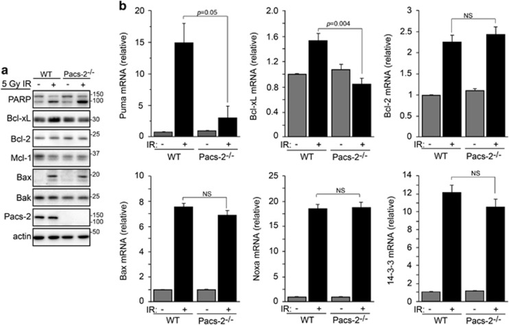
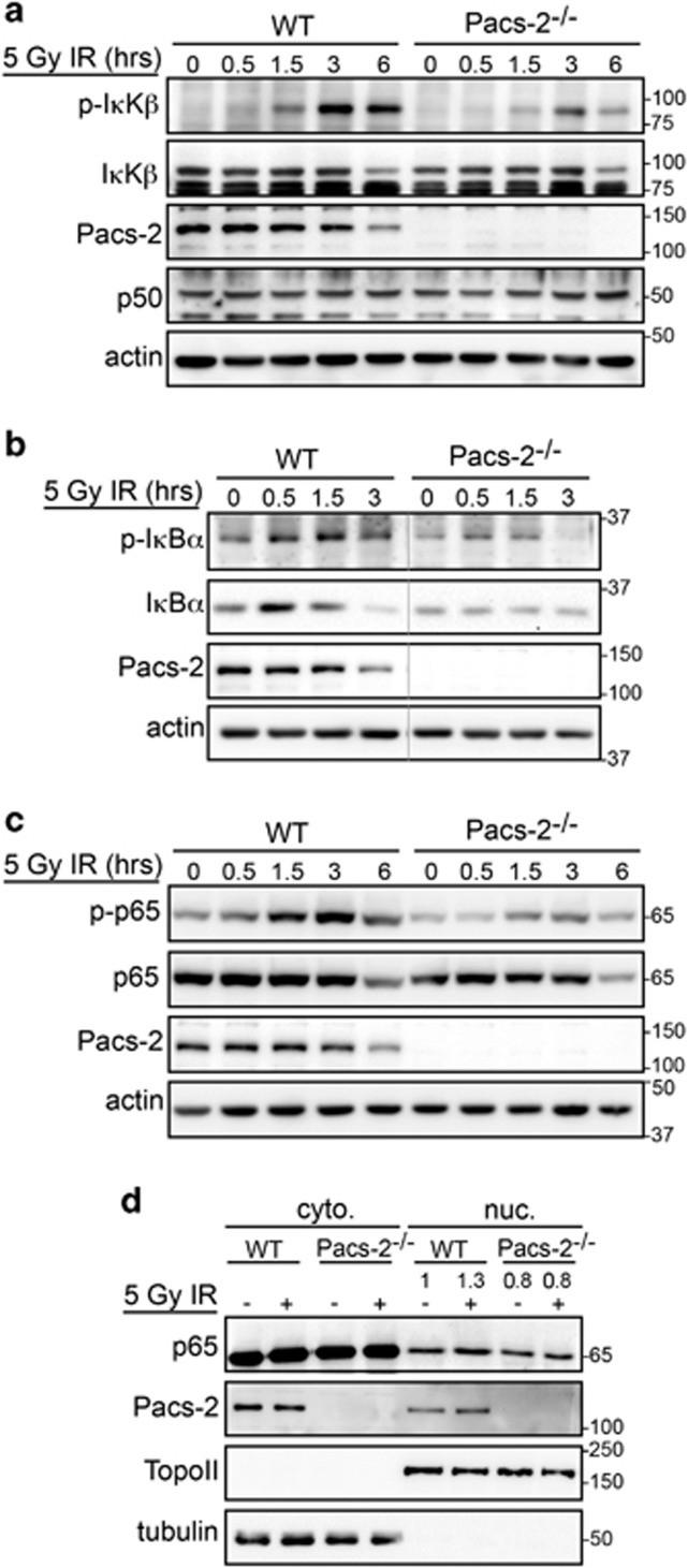
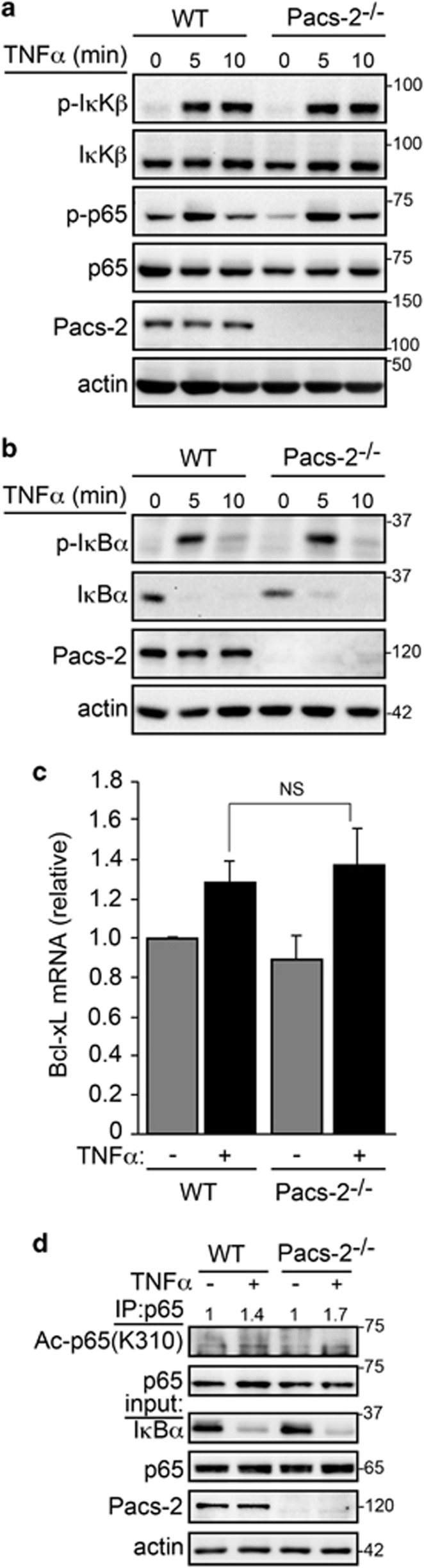
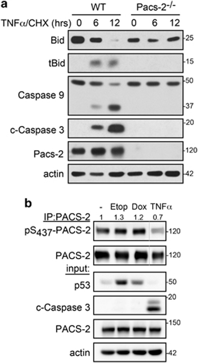
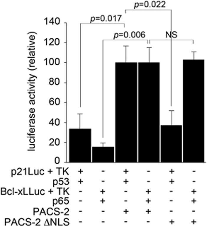
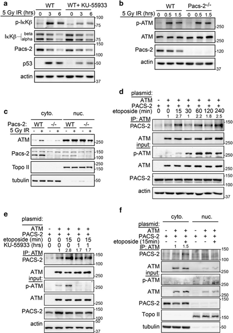
Nuclear factor kappa B (NF-κB) promotes cell survival in response to genotoxic stress by inducing the expression of anti-apoptotic proteins including Bcl-xL, which protects mitochondria from stress-induced mitochondrial outer membrane permeabilization (MOMP). Here we show that the multifunctional sorting protein Pacs-2 (phosphofurin acidic cluster sorting protein-2) is required for Bcl-xL induction following DNA damage in primary mouse thymocytes. Consequently, in response to DNA damage, Pacs-2(-/-) thymocytes exhibit a blunted induction of Bcl-xL, increased MOMP and accelerated apoptosis. Biochemical studies show that cytoplasmic PACS-2 promotes this DNA damage-induced anti-apoptotic pathway by interacting with ataxia telangiectasia mutated (ATM) to drive NF-κB activation and induction of Bcl-xL. However, Pacs-2 was not required for tumor necrosis factor-α-induced NF-κB activation, suggesting a role for PACS-2 selectively in NF-κB activation in response to DNA damage. These findings identify PACS-2 as an in vivo mediator of the ATM and NF-κB-dependent induction of Bcl-xL that promotes cell survival in response to DNA damage.


Figures

Figure 1
Figure 1
IR-induced apoptosis is accelerated in Pacs-2−/− thymocytes. (a) WT and Pacs-2−/− thymocytes were exposed or not to 4.5 Gy IR (6 h) and analyzed by flow cytometry for apoptotic cell death using propidium iodide/Annexin V co-staining. The frequency of early- and late apoptotic cells is shown in the lower right and upper right gates, respectively. (b) Thymocytes from WT and Pacs-2−/− mice were untreated or exposed to 5 Gy IR, harvested at the indicated times and cleaved caspase-3 measured by western blotting. Actin was used as loading control. (c) WT and Pacs-2−/− thymocytes were untreated or exposed to 5 Gy IR. At 4-h post IR, crude mitochondria and cytosol fractions were prepared and analyzed by western blotting for cytochrome c protein staining. Tubulin was used as control of cytoplasmic fraction loading
Figure 2
Figure 2
Pacs-2 depletion alters induction of pro- and anti-apoptotic targets genes. (a) WT and Pacs-2−/− thymocytes exposed or not to 5 Gy IR (4 h) were analyzed by western blotting for the induction of the indicated protein targets. PARP cleavage was used as a marker of increased apoptosis. Actin was used as loading control. (b) WT and Pacs-2−/− mice were exposed to 5 Gy WBI and isolated thymocytes were analyzed for induction of the indicated genes by qPCR (normalized to GAPDH). Error bars represent mean±S.E.M. from four or more mice per condition. Statistical significance was determined using Student's t-test
Figure 3
Figure 3
Pacs-2 modulates NF-κB activation in vivo following DNA damage. (a) WT and Pacs-2−/− thymocytes were exposed to 5 Gy IR and analyzed at increasing times for the phosphorylation of IκKβ Ser176/180 by western blotting. Actin was used as loading control. (b) WT and Pacs-2−/− thymocytes were exposed to 5 Gy IR and analyzed at increasing times for the phosphorylation of IκBα at Ser32/36 and degradation (IκBα lane) by western blotting. Actin was used as protein loading control. The unspliced blots are shown in Supplementary Figure S5A. (c) Thymuses from WT and Pacs-2−/− mice were exposed to 5 Gy IR and analyzed at increasing times for the phosphorylation of pSer536-p65 by western blotting. (d) WT and Pacs-2−/− thymocytes were untreated or exposed to 5 Gy IR. At 4-h post IR, nuclear and cytosolic fractions were prepared and analyzed by western blotting for p65 distribution. Topo II and tubulin were used as markers for the nuclear and cytoplasmic fractions, respectively. p65 was quantified using AlphaView (ProteinSimple)
Figure 4
Figure 4
Pacs-2 is not required for canonical NF-κB activation. (a) WT and Pacs-2−/− thymocytes were treated with 20ng/ml TNFα and analyzed at increasing times for the phosphorylation of pSer176/180-IκKβ and pSer536-p65 by western blotting. Actin was used as protein loading control. (b) WT and Pacs-2−/− thymocytes were treated with 20ng/ml TNFα and analyzed at increasing times for the phosphorylation (pSer32/36) and degradation of IκBα by western blotting. Actin was used as the loading control. (c) WT and Pacs-2−/− thymocytes were treated with TNFα (20 ng/ml) for 30 min and analyzed for Bcl-xL induction by qPCR (normalized to GAPDH). Error bars represent mean±S.E.M. from three mice per condition. Statistical significance was determined using Student's t-test. (d) WT and Pacs-2−/− thymocytes were treated with TNFα (20 ng/ml) for 20 min. p65 was immunoprecipitated (IP) from whole-cell lysates, and Ac-Lys310-p65 and IκBα were analyzed by western blotting. Ac-Lys310-p65 was quantified using AlphaView (ProteinSimple)
Figure 5
Figure 5
TNFα, but not DNA, damage switches PACS-2 to become an apoptotic effector. (a) WT and Pacs-2−/− MEF cells were treated with TNFα (100 ng/ml) and cycloheximide (1 μg/ml) for the indicated times and lysates were analyzed by western blotting. (b) PACS-2-overexpressing HCT116 cells were treated with etoposide (50 μM), doxorubicin (1 μM) or human TNFα (40 ng/ml) as indicated. PACS-2 was immunoprecipitated (IP) and pSer437-PACS-2 was detected by western blotting. pSer437-PACS-2 was quantified using AlphaView (ProteinSimple). The PACS-2 and actin Mr values were determined using the MW standards. The unspliced blots are shown in Supplementary Figure S5B
Figure 6
Figure 6
Cytoplasmic PACS-2 mediates Bcl-xL reporter expression. HCT116 cells were transfected with a luciferase expression plasmid under the control of p21 or Bcl-xL promoter together with PACS-2 or PACS-2ΔNLS and the transcription factor p53 or p65. Cells were harvested 24 h after transfection and processed for luciferase activity. Luciferase values were normalized between samples using the signal from the co-transfected Renilla plasmid. Error bars represent mean±S.E.M. from at least three independent experiments. Statistical significance was determined using Student's t-test
Figure 7
Figure 7
DNA damage-induced interaction between ATM and PACS-2. (a) WT and Pacs-2−/− thymocytes were left untreated or pre-treated with ATM inhibitor KU-55933 for 1 h and then exposed to 5 Gy IR. Then, thymocytes were analyzed at increasing times for pSer176/180-IκKβ by western blotting. Actin was used as the loading control. Quantification of the Pacs-2 signal (AlphaView, ProteinSimple) revealed a 50% decrease in total Pacs-2 by 6-h post IR. (b) WT and Pacs-2−/− thymocytes were exposed to 5 Gy IR and analyzed at increasing times for pSer1987-ATM by western blotting. Actin was used as the loading control. (c) WT and Pacs-2−/− thymocytes were left untreated or exposed to 5 Gy IR. At 2-h post IR, nuclear and cytosolic fractions were prepared and ATM was detected by western blotting. Topo II and tubulin were used as nuclear and cytoplasmic markers, respectively. (d) HCT116 cells co-expressing PACS-2 and ATM were treated with etoposide (50 μM) for the indicated times. ATM was immunoprecipitated (IP), and co-precipitating PACS-2 was detected by western blotting. PACS-2 was quantified using AlphaView (ProteinSimple). Actin was used as loading control. (e) HCT116 cells co-expressing PACS-2 and ATM were treated with etoposide (50 μM) or KU-55933 or both drugs. ATM was IP and PACS-2 was detected by western blotting. PACS-2 was quantified using AlphaView (ProteinSimple). Actin was used as loading control. (f) HCT116 cells expressing PACS-2 or PACS-2/ATM were treated with or without etoposide (50 μM) as indicated, nuclear and cytoplasmic fractions were prepared, ATM was IP and co-precipitating PACS-2 was detected by western blotting. PACS-2 was quantified using AlphaView (ProteinSimple). Topo II and tubulin were used as markers for the nuclear and cytoplasmic fractions, respectively

Similar articles

Cited by

References

    1. Napetschnig J, Wu H. Molecular basis of NF-kappaB signaling. Annu Rev Biophys 2013; 42: 443–468. - PMC - PubMed
    1. Chen C, Edelstein LC, Gelinas C. The Rel/NF-kappaB family directly activates expression of the apoptosis inhibitor Bcl-x(L). Mol Cell Biol. 2000; 20: 2687–2695. - PMC - PubMed
    1. Thomenius MJ, Distelhorst CW. Bcl-2 on the endoplasmic reticulum: protecting the mitochondria from a distance. J Cell Sci 2003; 116(Pt 22): 4493–4499. - PubMed
    1. Eno CO, Eckenrode EF, Olberding KE, Zhao G, White C, Li C. Distinct roles of mitochondria- and ER-localized Bcl-xL in apoptosis resistance and Ca2+ homeostasis. Mol Biol Cell 2012; 23: 2605–2618. - PMC - PubMed
    1. Billen LP, Kokoski CL, Lovell JF, Leber B, Andrews DW. Bcl-XL inhibits membrane permeabilization by competing with Bax. PLoS Biol. 2008; 6: e147. - PMC - PubMed

Publication types

MeSH terms